文献检索文档翻译深度研究
Suppr Zotero 插件Zotero 插件
邀请有礼套餐&价格历史记录

新学期,新优惠

限时优惠:9月1日-9月22日

30天高级会员仅需29元

1天体验卡首发特惠仅需5.99元

了解详情
不再提醒
插件&应用
Suppr Zotero 插件Zotero 插件浏览器插件Mac 客户端Windows 客户端微信小程序
高级版
套餐订阅购买积分包
AI 工具
文献检索文档翻译深度研究
关于我们
关于 Suppr公司介绍联系我们用户协议隐私条款
关注我们

Suppr 超能文献

核心技术专利:CN118964589B侵权必究
粤ICP备2023148730 号-1Suppr @ 2025

应用磁粒子成像技术进行保乳手术术中切缘分析的构想。

Concept for using magnetic particle imaging for intraoperative margin analysis in breast-conserving surgery.

机构信息

Department of Radiology, A. A. Martinos Center for Biomedical Imaging, Massachusetts General Hospital, Charlestown, MA, 02129, USA.

Harvard-MIT Division of Health Sciences and Technology, Cambridge, MA, 02139, USA.

出版信息

Sci Rep. 2021 Jun 29;11(1):13456. doi: 10.1038/s41598-021-92644-8.


DOI:10.1038/s41598-021-92644-8
PMID:34188077
原文链接:https://pmc.ncbi.nlm.nih.gov/articles/PMC8242088/
Abstract

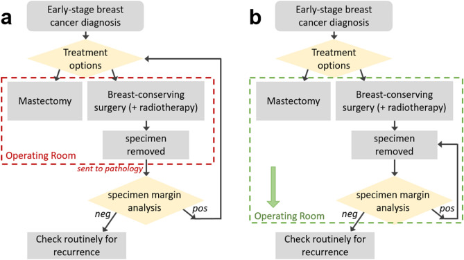
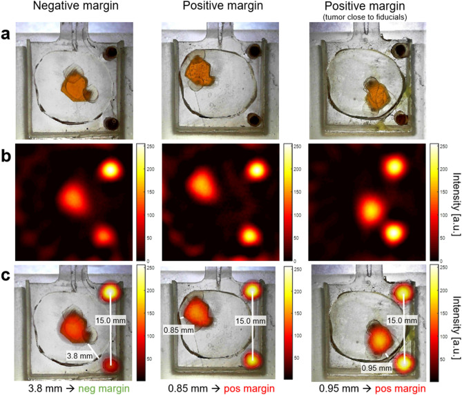
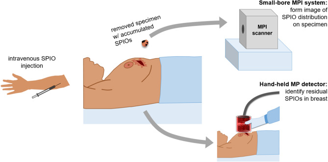
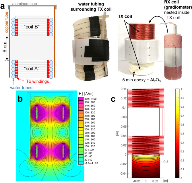
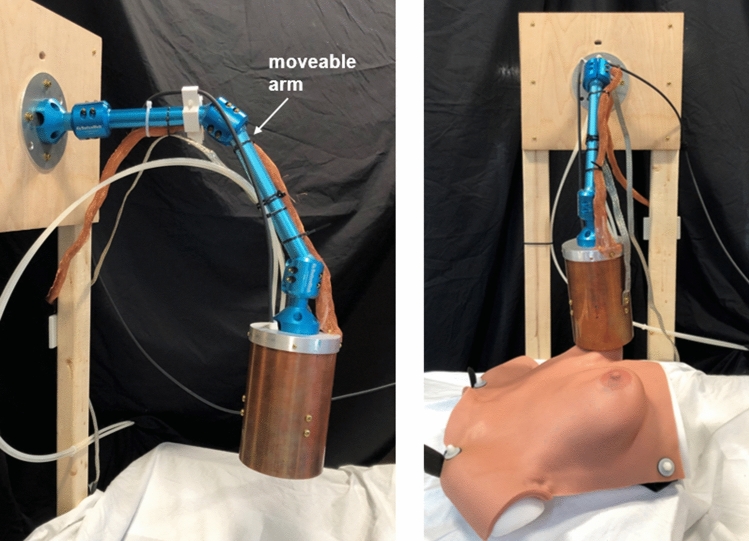
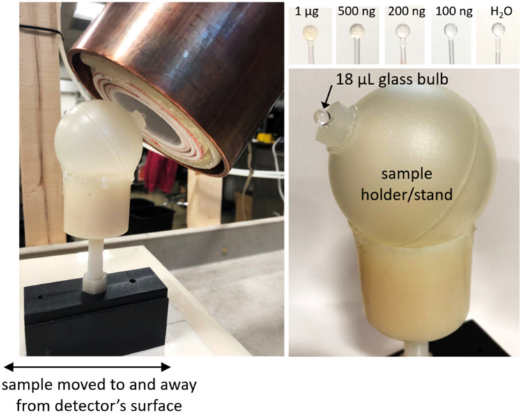
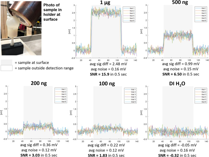
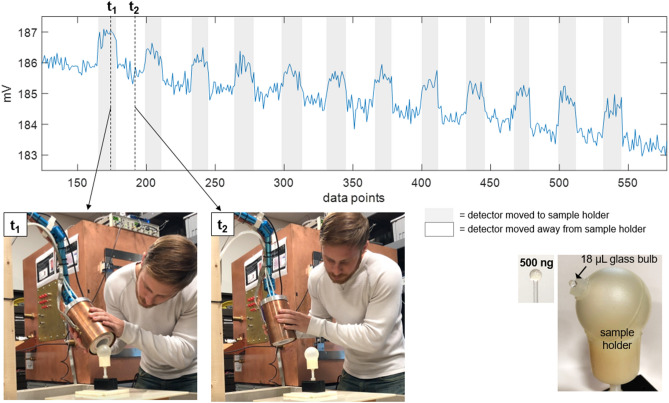
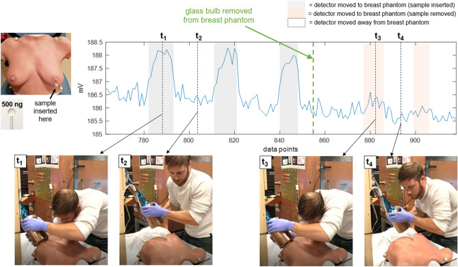
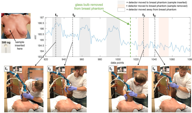
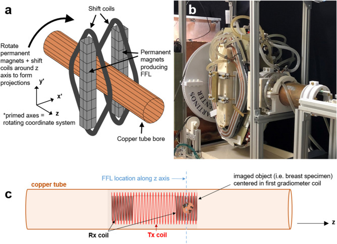
Breast-conserving surgery (BCS) is a commonly utilized treatment for early stage breast cancers but has relatively high reexcision rates due to post-surgical identification of positive margins. A fast, specific, sensitive, easy-to-use tool for assessing margins intraoperatively could reduce the need for additional surgeries, and while many techniques have been explored, the clinical need is still unmet. We assess the potential of Magnetic Particle Imaging (MPI) for intraoperative margin assessment in BCS, using a passively or actively tumor-targeted iron oxide agent and two hardware devices: a hand-held Magnetic Particle detector for identifying residual tumor in the breast, and a small-bore MPI scanner for quickly imaging the tumor distribution in the excised specimen. Here, we present both hardware systems and demonstrate proof-of-concept detection and imaging of clinically relevant phantoms.

摘要

保乳手术(BCS)是一种常用于早期乳腺癌的治疗方法,但由于术后发现阳性边缘,其再次切除率相对较高。一种快速、特异、敏感、易于使用的术中评估切缘的工具可以减少额外手术的需要,尽管已经探索了许多技术,但临床需求仍未得到满足。我们评估了磁性粒子成像(MPI)在保乳手术中用于术中切缘评估的潜力,使用了一种被动或主动肿瘤靶向氧化铁剂和两种硬件设备:一种手持式磁性粒子探测器,用于识别乳房中的残留肿瘤,以及一种小口径 MPI 扫描仪,用于快速成像切除标本中的肿瘤分布。在这里,我们展示了这两种硬件系统,并证明了临床相关模型的探测和成像的概念验证。

https://cdn.ncbi.nlm.nih.gov/pmc/blobs/05ce/8242088/5fe2ae9b07ed/41598_2021_92644_Fig11_HTML.jpg
https://cdn.ncbi.nlm.nih.gov/pmc/blobs/05ce/8242088/e5c7195783eb/41598_2021_92644_Fig1_HTML.jpg
https://cdn.ncbi.nlm.nih.gov/pmc/blobs/05ce/8242088/2c2bb1d2e754/41598_2021_92644_Fig2_HTML.jpg
https://cdn.ncbi.nlm.nih.gov/pmc/blobs/05ce/8242088/93966fc51aac/41598_2021_92644_Fig3_HTML.jpg
https://cdn.ncbi.nlm.nih.gov/pmc/blobs/05ce/8242088/d97c7a088ac7/41598_2021_92644_Fig4_HTML.jpg
https://cdn.ncbi.nlm.nih.gov/pmc/blobs/05ce/8242088/7355a4da0640/41598_2021_92644_Fig5_HTML.jpg
https://cdn.ncbi.nlm.nih.gov/pmc/blobs/05ce/8242088/7e17ffbefdba/41598_2021_92644_Fig6_HTML.jpg
https://cdn.ncbi.nlm.nih.gov/pmc/blobs/05ce/8242088/44e1bf288088/41598_2021_92644_Fig7_HTML.jpg
https://cdn.ncbi.nlm.nih.gov/pmc/blobs/05ce/8242088/2215768d5bcf/41598_2021_92644_Fig8_HTML.jpg
https://cdn.ncbi.nlm.nih.gov/pmc/blobs/05ce/8242088/0553a3189385/41598_2021_92644_Fig9_HTML.jpg
https://cdn.ncbi.nlm.nih.gov/pmc/blobs/05ce/8242088/04138ee7d5a4/41598_2021_92644_Fig10_HTML.jpg
https://cdn.ncbi.nlm.nih.gov/pmc/blobs/05ce/8242088/5fe2ae9b07ed/41598_2021_92644_Fig11_HTML.jpg

相似文献

[1]
Concept for using magnetic particle imaging for intraoperative margin analysis in breast-conserving surgery.

Sci Rep. 2021-6-29

[2]
Intraoperative Assessment of Tumor Resection Margins in Breast-Conserving Surgery Using F-FDG Cerenkov Luminescence Imaging: A First-in-Human Feasibility Study.

J Nucl Med. 2016-12-8

[3]
Biophotonic technologies for assessment of breast tumor surgical margins-A review.

J Biophotonics. 2021-1

[4]
Imaging of lumpectomy surface with large field-of-view confocal laser scanning microscope for intraoperative margin assessment - POLARHIS study.

Breast. 2022-12

[5]
Clinicopathologic factors related to surgical margin involvement, reoperation, and residual cancer in primary operable breast cancer - An analysis of 2050 patients.

Eur J Surg Oncol. 2018-8-1

[6]
Definition and Management of Positive Margins for Invasive Breast Cancer.

Surg Clin North Am. 2018-4-24

[7]
Diagnostic Accuracy of Quantitative Micro-Elastography for Margin Assessment in Breast-Conserving Surgery.

Cancer Res. 2020-4-15

[8]
An intraoperative MRI system for margin assessment in breast conserving surgery: Initial results from a novel technique.

J Surg Oncol. 2016-7

[9]
Near-infrared fluorescence (NIRF) imaging in breast-conserving surgery: assessing intraoperative techniques in tissue-simulating breast phantoms.

Eur J Surg Oncol. 2010-11-24

[10]
Clinical Impact of Intraoperative Margin Assessment in Breast-Conserving Surgery With a Novel Pegulicianine Fluorescence-Guided System: A Nonrandomized Controlled Trial.

JAMA Surg. 2022-7-1

引用本文的文献

[1]
Principles and applications of magnetic nanomaterials in magnetically guided bioimaging.

Mater Today Phys. 2023-3

[2]
Assessing the efficacy of intraoperative fluorescence imaging for tumor margin identification in breast conserving surgery.

Bioinformation. 2024-12-31

[3]
Development of Iron Oxide Nanochains as a Sensitive Magnetic Particle Imaging Tracer for Cancer Detection.

ACS Appl Mater Interfaces. 2025-4-9

[4]
Magnetic particle imaging angiography of the femoral artery in a human cadaveric perfusion model.

Commun Med (Lond). 2025-3-13

[5]
Non-FFP-Based Magnetic Particle Imaging (NFMPI) with an Open-Type RF Coil System: A Feasibility Study.

Sensors (Basel). 2025-1-23

[6]
Foundational insights for theranostic applications of magnetoelectric nanoparticles.

Nanoscale Horiz. 2025-3-24

[7]
Tomographic Magnetic Particle Imaging With a Single-Sided Field-Free Line Scanner.

IEEE Trans Biomed Eng. 2024-12

[8]
Harmonic dependence of thermal magnetic particle imaging.

Sci Rep. 2023-9-22

[9]
Implementation of the surface gradiometer receive coils for the improved detection limit and sensitivity in the single-sided MPI scanner.

Phys Med Biol. 2022-12-9

[10]
Recent developments of the reconstruction in magnetic particle imaging.

Vis Comput Ind Biomed Art. 2022-10-1

本文引用的文献

[1]
The Applications of Magnetic Particle Imaging: From Cell to Body.

Diagnostics (Basel). 2020-10-9

[2]
Hybrid MPI-MRI System for Dual-Modal In Situ Cardiovascular Assessments of Real-Time 3D Blood Flow Quantification-A Pre-Clinical In Vivo Feasibility Investigation.

IEEE Trans Med Imaging. 2020-12

[3]
Tomographic Field Free Line Magnetic Particle Imaging With an Open-Sided Scanner Configuration.

IEEE Trans Med Imaging. 2020-12

[4]
Using magnetic particle imaging systems to localize and guide magnetic hyperthermia treatment: tracers, hardware, and future medical applications.

Theranostics. 2020

[5]
Three-dimensional surface imaging in breast cancer: a new tool for clinical studies?

Radiat Oncol. 2020-2-28

[6]
Magnetic Particle Imaging of Macrophages Associated with Cancer: Filling the Voids Left by Iron-Based Magnetic Resonance Imaging.

Mol Imaging Biol. 2020-8

[7]
The entry of nanoparticles into solid tumours.

Nat Mater. 2020-1-13

[8]
Magnetic particle imaging in vascular medicine.

Innov Surg Sci. 2018-10-9

[9]
A prospective, single-arm, multicentre clinical evaluation of a new localisation technique using non-radioactive Magseeds for surgery of clinically occult breast lesions.

Clin Radiol. 2019-9-25

[10]
Magnetic Particle Imaging meets Computed Tomography: first simultaneous imaging.

Sci Rep. 2019-9-2

文献AI研究员

20分钟写一篇综述,助力文献阅读效率提升50倍

立即体验

用中文搜PubMed

大模型驱动的PubMed中文搜索引擎

马上搜索

推荐工具

医学文档翻译智能文献检索