Rispoli Marco, Eandi Chiara M, Di Antonio Luca, Kilian Raphael, Montesel Andrea, Savastano Maria C
Chorioretinal Vasculopathies Unit, Surgery and Emergency Ophthalmology Department, Eye Hospital, 00136 Rome, Italy.
Department of Ophthalmology, Jules Gonin Eye Hospital, Fondation Asile des Aveugles, University of Lausanne, 1002 Lausanne, Switzerland.
Biomedicines. 2021 Jun 10;9(6):668. doi: 10.3390/biomedicines9060668.
The purpose of this study was to describe early changes in the morphology of pigment epithelium detachments (PED) after an intravitreal injection of Brolucizumab into eyes with macular neovascularization secondary to exudative age-related macular degeneration (e-AMD).
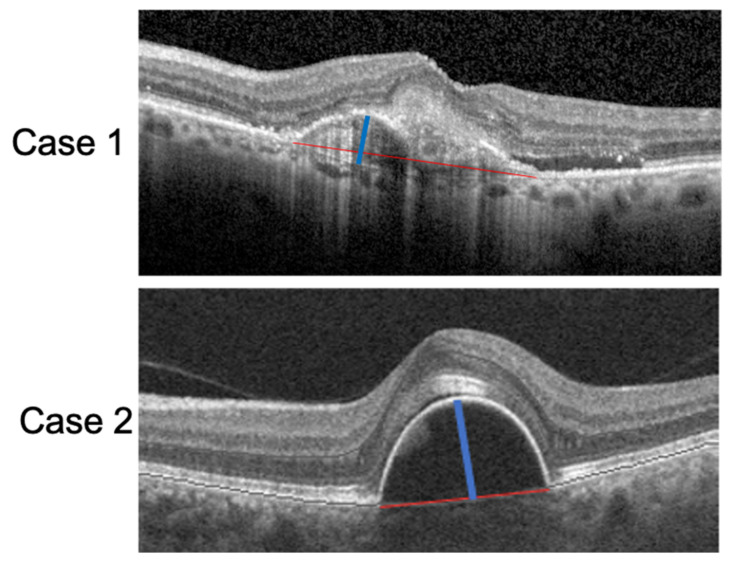
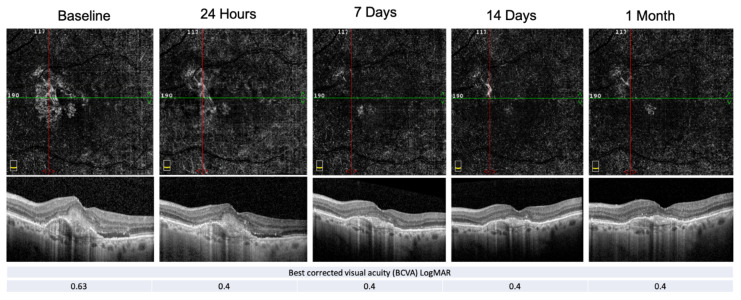
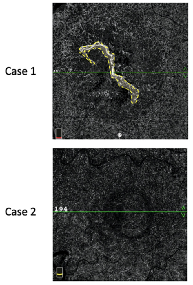
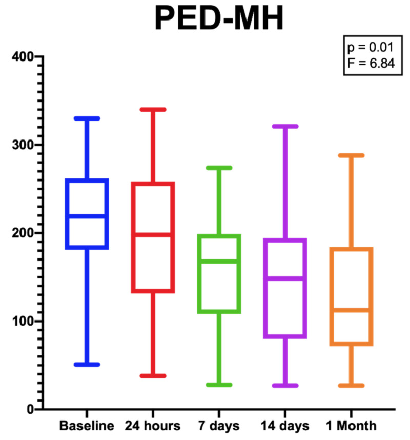
We included twelve eyes of 12 patients with PED secondary to e-AMD which were not responding to prior anti-VEGF treatments. An ophthalmic examination and an assessment of PED-horizontal maximal diameter (PED-HMD), PED-maximum high (PED-MH) and macular neovascularization (MNV) flow area (MNV-FA) by the means of structural optical coherence tomography (OCT) and OCT Angiography (OCT-A) were performed at baseline, as well as 1, 7, 14 and 30 days after the injection.
The mean age of the population of study was 78.4 (SD ± 4.8). The mean number of previous Ranibizumab or Aflibercept injections was 13 (SD ± 8). At the last follow-up visit, the PED-HMD did not significantly change ( = 0.16; F(DF:1.94, 20,85) = 1.9), the PED-MH showed a significant reduction [ = 0.01; F(DF:1.31, 14.13) = 6.84.] and the MNV-FA did not significantly differ ( = 0.1; F(1.97, 21.67) = 2.54) from baseline. No signs of ocular inflammation were observed during follow-up.
A single Brolucizumab injection was able to determine the short-term effects on PEDs' anatomical features of eyes with an unresponsive e-AMD.
本研究的目的是描述在玻璃体内注射布罗珠单抗后,继发于渗出性年龄相关性黄斑变性(e-AMD)的黄斑新生血管眼色素上皮脱离(PED)形态的早期变化。
我们纳入了12例继发于e-AMD且对先前抗VEGF治疗无反应的PED患者的12只眼。在基线以及注射后1天、7天、14天和30天,通过结构光学相干断层扫描(OCT)和OCT血管造影(OCT-A)进行眼科检查,并评估PED水平最大直径(PED-HMD)、PED最大高度(PED-MH)和黄斑新生血管(MNV)血流面积(MNV-FA)。
研究人群的平均年龄为78.4(标准差±4.8)。先前雷珠单抗或阿柏西普注射的平均次数为13次(标准差±8)。在最后一次随访时,PED-HMD没有显著变化(P = 0.16;F(自由度:1.94,20.85)= 1.9),PED-MH显示出显著降低[P = 0.01;F(自由度:1.31,14.13)= 6.84],并且MNV-FA与基线相比没有显著差异(P = 0.1;F(1.97,21.67)= 2.54)。随访期间未观察到眼部炎症迹象。
单次注射布罗珠单抗能够确定对无反应的e-AMD眼PED解剖特征的短期影响。