Umutlu Lale, Kirchner Julian, Bruckmann Nils Martin, Morawitz Janna, Antoch Gerald, Ingenwerth Marc, Bittner Ann-Kathrin, Hoffmann Oliver, Haubold Johannes, Grueneisen Johannes, Quick Harald H, Rischpler Christoph, Herrmann Ken, Gibbs Peter, Pinker-Domenig Katja
Department of Diagnostic and Interventional Radiology and Neuroradiology, University Hospital Essen, University of Duisburg-Essen, D-45147 Essen, Germany.
Department of Radiology, Memorial Sloan Kettering Cancer Center, New York, NY 10065, USA.
Cancers (Basel). 2021 Jun 11;13(12):2928. doi: 10.3390/cancers13122928.
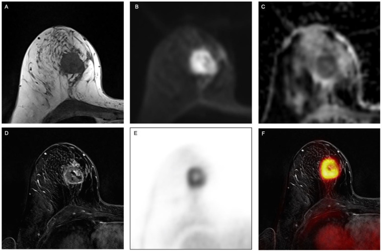
This study investigated the performance of simultaneous F-FDG PET/MRI of the breast as a platform for comprehensive radiomics analysis for breast cancer subtype analysis, hormone receptor status, proliferation rate and lymphonodular and distant metastatic spread.
One hundred and twenty-four patients underwent simultaneous F-FDG PET/MRI. Breast tumors were segmented and radiomic features were extracted utilizing CERR software following the IBSI guidelines. LASSO regression was employed to select the most important radiomics features prior to model development. Five-fold cross validation was then utilized alongside support vector machines, resulting in predictive models for various combinations of imaging data series.
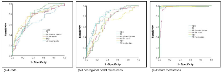
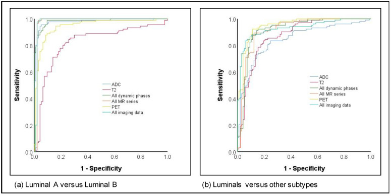
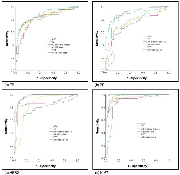
The highest AUC and accuracy for differentiation between luminal A and B was achieved by all MR sequences (AUC 0.98; accuracy 97.3). The best results in AUC for prediction of hormone receptor status and proliferation rate were found based on all MR and PET data (ER AUC 0.87, PR AUC 0.88, Ki-67 AUC 0.997). PET provided the best determination of grading (AUC 0.71), while all MR and PET analyses yielded the best results for lymphonodular and distant metastatic spread (0.81 and 0.99, respectively).
F-FDG PET/MRI enables comprehensive high-quality radiomics analysis for breast cancer phenotyping and tumor decoding, utilizing the perks of simultaneously acquired morphologic, functional and metabolic data.
本研究调查了乳腺同时进行F-FDG PET/MRI作为乳腺癌亚型分析、激素受体状态、增殖率以及淋巴结和远处转移扩散综合放射组学分析平台的性能。
124例患者接受了同时进行的F-FDG PET/MRI检查。利用CERR软件按照IBSI指南对乳腺肿瘤进行分割并提取放射组学特征。在模型开发之前,采用LASSO回归选择最重要的放射组学特征。然后使用五折交叉验证和支持向量机,生成针对各种成像数据系列组合的预测模型。
所有MR序列在区分管腔A型和B型方面的AUC和准确性最高(AUC 0.98;准确性97.3)。基于所有MR和PET数据,在预测激素受体状态和增殖率的AUC方面取得了最佳结果(ER AUC 0.87,PR AUC 0.88,Ki-67 AUC 0.997)。PET在分级判定方面表现最佳(AUC 0.71),而所有MR和PET分析在淋巴结和远处转移扩散方面取得了最佳结果(分别为0.81和0.99)。
F-FDG PET/MRI利用同时获取的形态学、功能和代谢数据的优势,能够对乳腺癌进行全面的高质量放射组学分析,以实现乳腺癌表型分析和肿瘤解码。