Jia Yanlu, Liu Chunling, Li Hui, Li Xiaonan, Wu Jun, Zhao Yimin, Xu Mengya, Yu Haitao, Guan Zhitong, Sun Shuning, Zhang Chao, Duan Zhiyi
Department of Neurology, The Second Affiliated Hospital of Zhengzhou University, Zhengzhou City, Henan Province, 450000, People's Republic of China.
Nat Sci Sleep. 2021 Jun 25;13:863-872. doi: 10.2147/NSS.S305465. eCollection 2021.
There is increasing evidence of a causal interaction between obstructive sleep apnea (OSA) and white matter hyperintensity (WMH). WMH and enlarged perivascular space (EPVS) are the neuroimaging markers for cerebral small vessel disease (CSVD). Thus, this study aimed to determine whether a contextual relationship existed between OSA and EPVS.
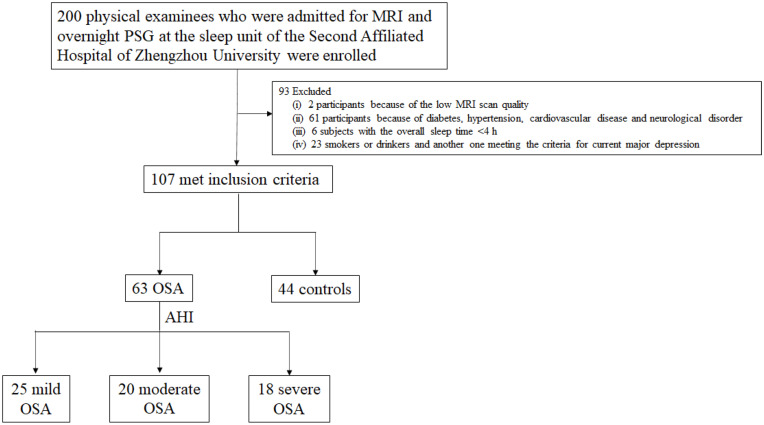
In this study, 107 participants underwent 1-night polysomnography, brain magnetic resonance imaging (MRI) and health screening examinations and were classified as 63 OSA patients (mild, moderate, and severe groups), and 44 healthy controls. We assessed the sleep characteristics in OSA group, quantified the total EPVS from MRI and related them to the measures of polysomnography-obtained sleep parameters.
Polysomnography revealed that 63 OSA patients had sleep architecture alteration. A higher proportion of N2 phase sleep (N2%), lower percentage of N3 sleep (N3%) and REM sleep (REM%), as well as increased arousal index (AI), oxygen desaturation index (ODI) and decreased lowest arterial oxygen saturation (LSaO2) were detected. The results also indicated a higher prevalence and a larger number of EPVS, and a lower Mini Mental State Scale (MMSE) scale score in OSA group. LSaO2, N3% and REM% were negatively correlated with the total EPVS, whereas ODI, AI and N2% were positively correlated with the total EPVS.
The findings suggested that OSA patients had sleep disturbances with a higher incidence and more severe EPVS. Furthermore, the EPVS in OSA might be secondary to sleep disturbances, intermittent hypoxemia and the respiratory event-related hemodynamic changes. Thus, our findings highlighted that increased risk for EPVS in OSA is a potential contributor to increased stroke risk in OSA.
越来越多的证据表明阻塞性睡眠呼吸暂停(OSA)与脑白质高信号(WMH)之间存在因果相互作用。WMH和血管周围间隙扩大(EPVS)是脑小血管疾病(CSVD)的神经影像学标志物。因此,本研究旨在确定OSA与EPVS之间是否存在背景关系。
在本研究中,107名参与者接受了1晚的多导睡眠图检查、脑磁共振成像(MRI)和健康筛查检查,并被分为63名OSA患者(轻度、中度和重度组)和44名健康对照者。我们评估了OSA组的睡眠特征,从MRI中量化了总的EPVS,并将其与多导睡眠图获得的睡眠参数测量值相关联。
多导睡眠图显示63名OSA患者存在睡眠结构改变。检测到N2期睡眠比例较高(N2%)、N3期睡眠(N3%)和快速眼动睡眠(REM%)比例较低,以及觉醒指数(AI)、氧饱和度下降指数(ODI)增加和最低动脉血氧饱和度(LSaO2)降低。结果还表明,OSA组中EPVS的患病率更高、数量更多,且简易精神状态检查表(MMSE)评分更低。LSaO2、N3%和REM%与总的EPVS呈负相关,而ODI、AI和N2%与总的EPVS呈正相关。
研究结果表明,OSA患者存在睡眠障碍,且EPVS的发生率更高、更严重。此外,OSA中的EPVS可能继发于睡眠障碍、间歇性低氧血症和与呼吸事件相关的血流动力学变化。因此,我们的研究结果强调,OSA中EPVS风险增加是OSA中风风险增加的一个潜在因素。