Department of Cellular and Molecular Biophysics, Max Planck Institute of Biochemistry, Martinsried, Germany.
Department of Molecular Structural Biology, Max Planck Institute of Biochemistry, Martinsried, Germany.
Nat Commun. 2021 Jul 2;12(1):4086. doi: 10.1038/s41467-021-24049-0.
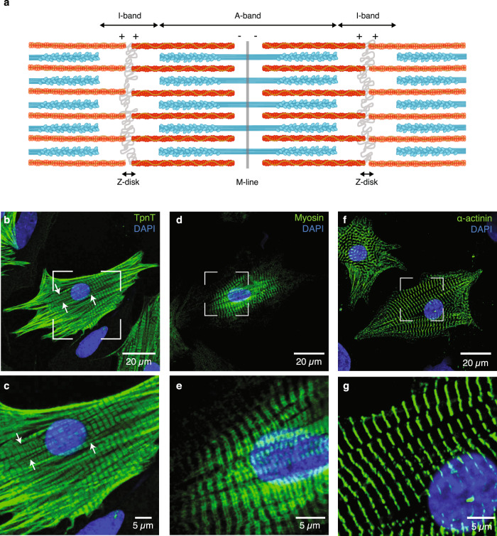
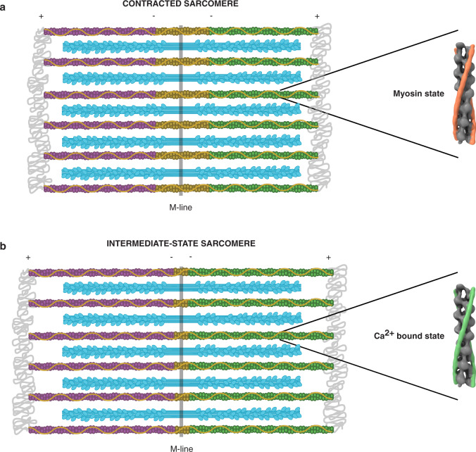
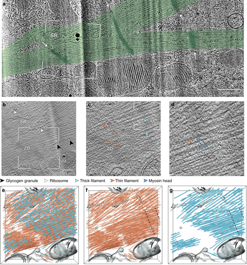
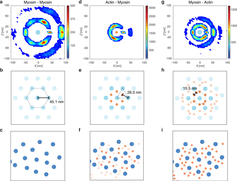
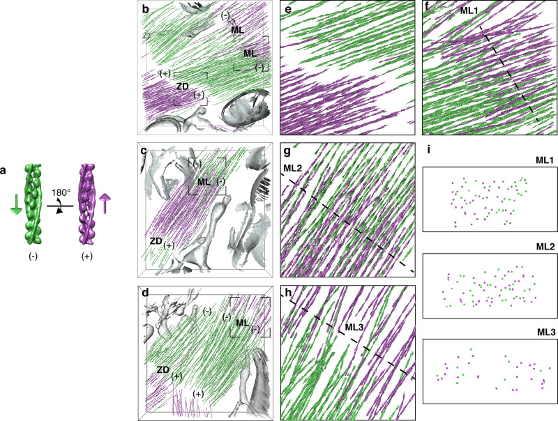
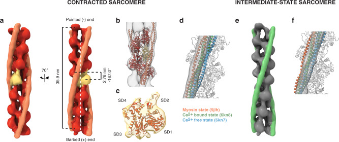
Sarcomeres, the basic contractile units of striated muscle, produce the forces driving muscular contraction through cross-bridge interactions between actin-containing thin filaments and myosin II-based thick filaments. Until now, direct visualization of the molecular architecture underlying sarcomere contractility has remained elusive. Here, we use in situ cryo-electron tomography to unveil sarcomere contraction in frozen-hydrated neonatal rat cardiomyocytes. We show that the hexagonal lattice of the thick filaments is already established at the neonatal stage, with an excess of thin filaments outside the trigonal positions. Structural assessment of actin polarity by subtomogram averaging reveals that thin filaments in the fully activated state form overlapping arrays of opposite polarity in the center of the sarcomere. Our approach provides direct evidence for thin filament sliding during muscle contraction and may serve as a basis for structural understanding of thin filament activation and actomyosin interactions inside unperturbed cellular environments.
肌节是横纹肌的基本收缩单位,通过肌动蛋白含有的细丝和肌球蛋白 II 为基础的粗丝之间的交联相互作用产生驱动肌肉收缩的力。到目前为止,肌节收缩的分子结构仍然难以直接可视化。在这里,我们使用原位冷冻电子断层扫描技术揭示了冷冻水合新生大鼠心肌细胞中的肌节收缩。我们表明,在新生儿期,粗丝的六方晶格已经建立,三角位置以外有过多的细丝。通过亚断层平均法对肌动蛋白极性的结构评估表明,在完全激活状态下,细丝在肌节中心形成相互重叠的相反极性排列。我们的方法为肌肉收缩过程中细丝滑动提供了直接证据,并可能为在未受干扰的细胞环境中理解细丝激活和肌球蛋白相互作用的结构提供基础。