• 文献检索
  • 文档翻译
  • 深度研究
  • 学术资讯
  • Suppr Zotero 插件Zotero 插件
  • 邀请有礼
  • 套餐&价格
  • 历史记录
应用&插件
Suppr Zotero 插件Zotero 插件浏览器插件Mac 客户端Windows 客户端微信小程序
定价
高级版会员购买积分包购买API积分包
服务
文献检索文档翻译深度研究API 文档MCP 服务
关于我们
关于 Suppr公司介绍联系我们用户协议隐私条款
关注我们

Suppr 超能文献

核心技术专利:CN118964589B侵权必究
粤ICP备2023148730 号-1Suppr @ 2026

文献检索

告别复杂PubMed语法,用中文像聊天一样搜索,搜遍4000万医学文献。AI智能推荐,让科研检索更轻松。

立即免费搜索

文件翻译

保留排版,准确专业,支持PDF/Word/PPT等文件格式,支持 12+语言互译。

免费翻译文档

深度研究

AI帮你快速写综述,25分钟生成高质量综述,智能提取关键信息,辅助科研写作。

立即免费体验

释出-捕获机制在管状芽生过程中产生一个必需的非中心体微管阵列。

A release-and-capture mechanism generates an essential non-centrosomal microtubule array during tube budding.

机构信息

MRC-Laboratory of Molecular Biology, Cambridge, UK.

Wellcome-MRC Stem Cell Institute, Jeffrey Cheah Biomedical Centre, University of Cambridge, Cambridge, UK.

出版信息

Nat Commun. 2021 Jul 2;12(1):4096. doi: 10.1038/s41467-021-24332-0.

DOI:10.1038/s41467-021-24332-0
PMID:34215746
原文链接:https://pmc.ncbi.nlm.nih.gov/articles/PMC8253823/
Abstract

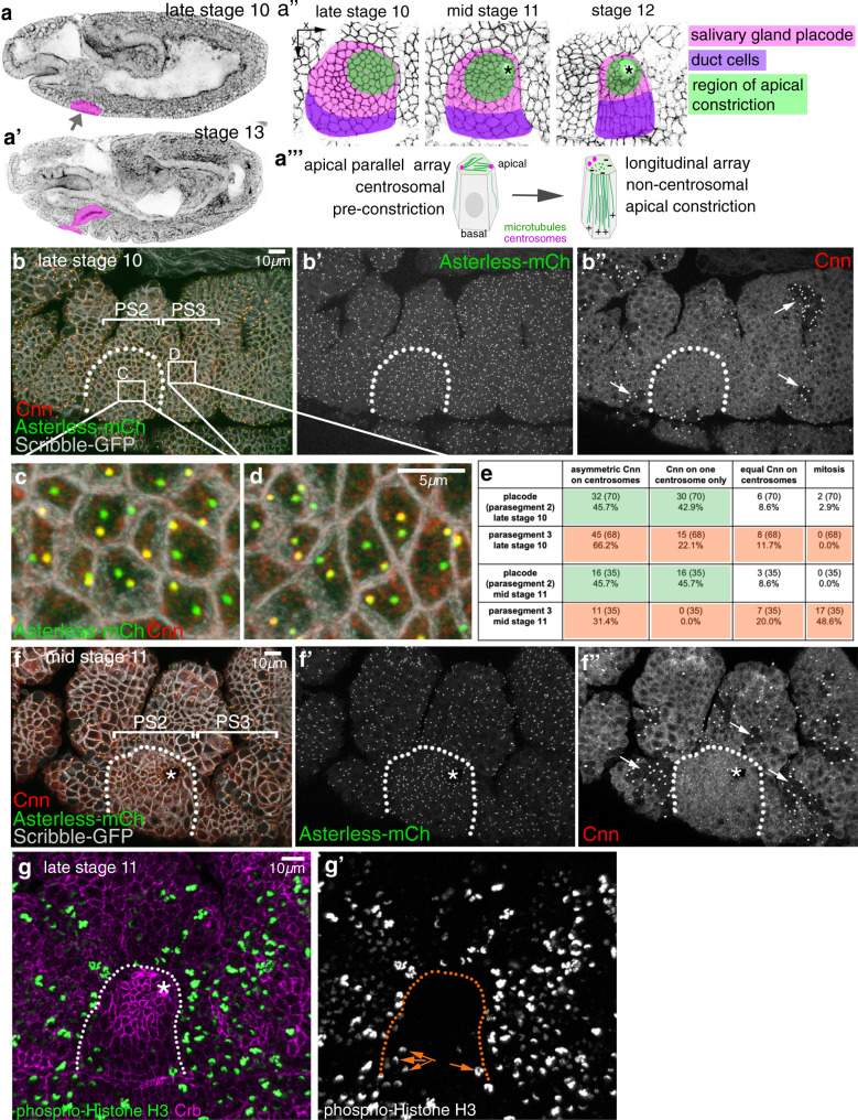
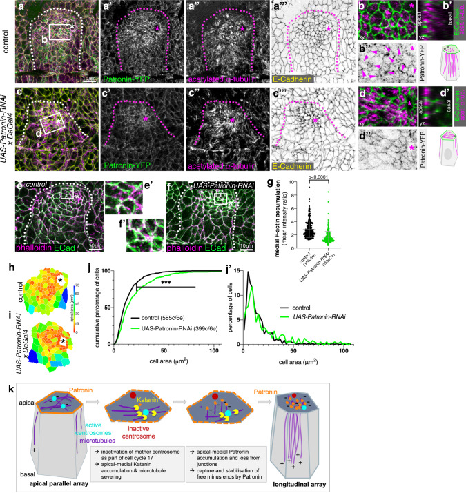
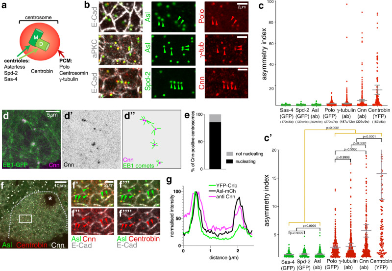
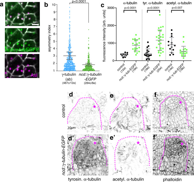
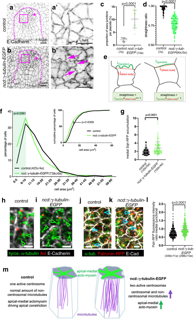
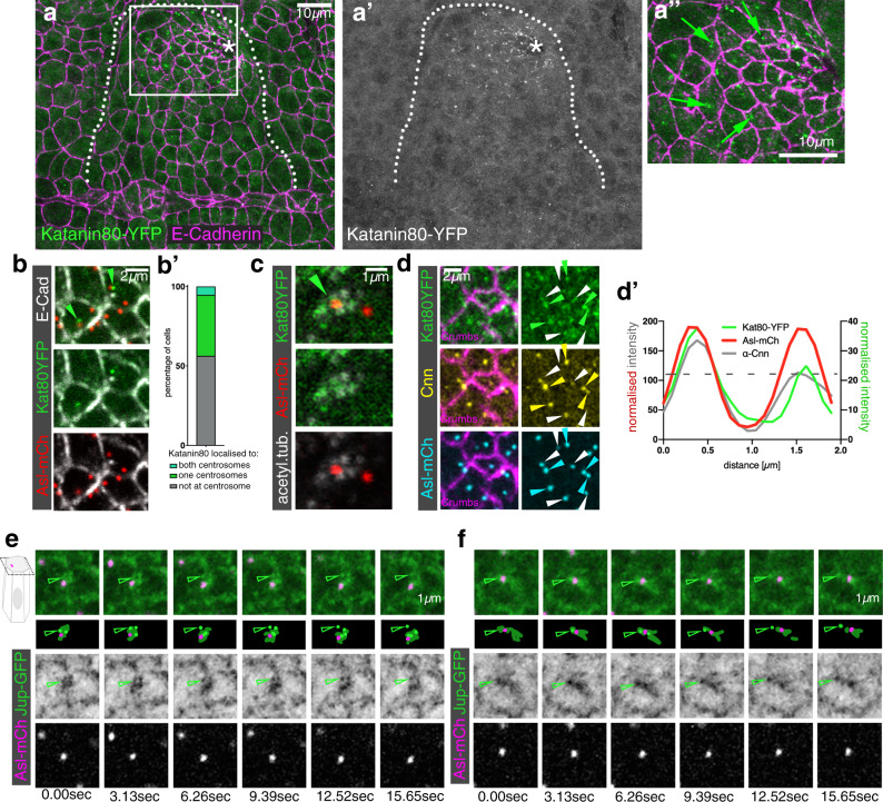
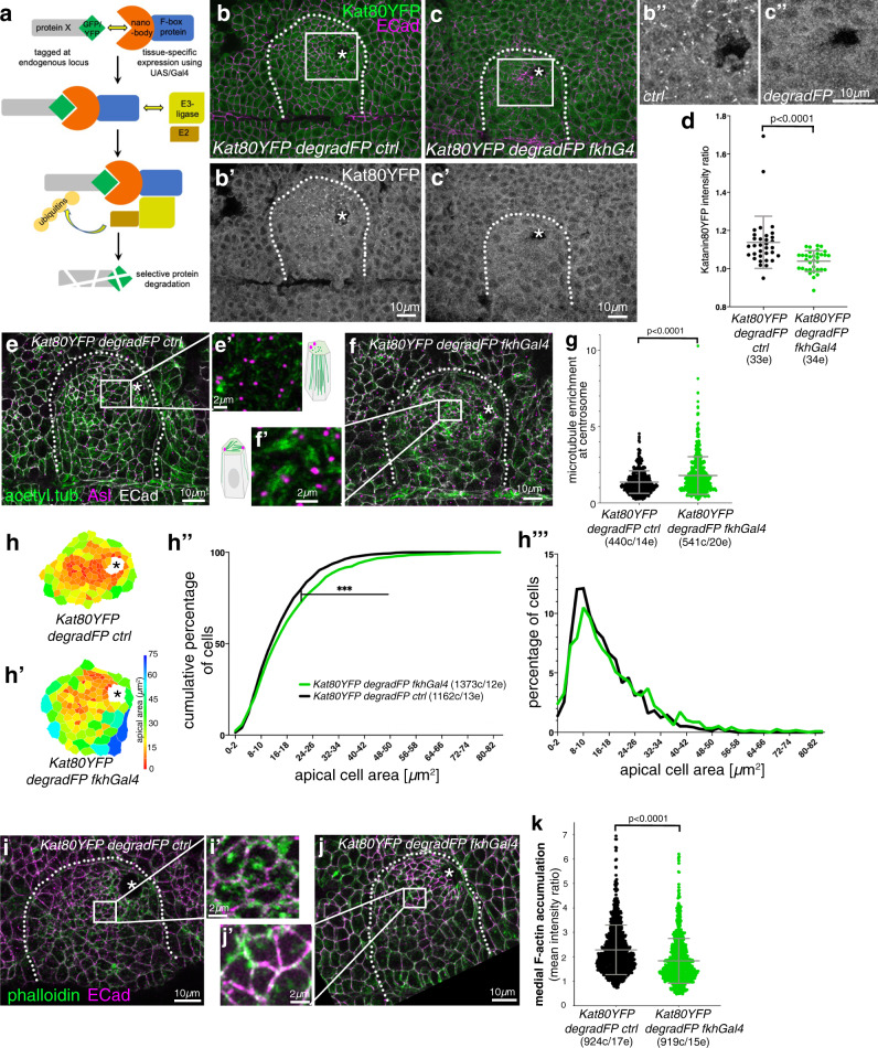
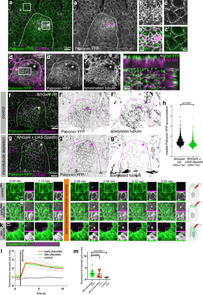
Non-centrosomal microtubule arrays serve crucial functions in cells, yet the mechanisms of their generation are poorly understood. During budding of the epithelial tubes of the salivary glands in the Drosophila embryo, we previously demonstrated that the activity of pulsatile apical-medial actomyosin depends on a longitudinal non-centrosomal microtubule array. Here we uncover that the exit from the last embryonic division cycle of the epidermal cells of the salivary gland placode leads to one centrosome in the cells losing all microtubule-nucleation capacity. This restriction of nucleation activity to the second, Centrobin-enriched, centrosome is key for proper morphogenesis. Furthermore, the microtubule-severing protein Katanin and the minus-end-binding protein Patronin accumulate in an apical-medial position only in placodal cells. Loss of either in the placode prevents formation of the longitudinal microtubule array and leads to loss of apical-medial actomyosin and impaired apical constriction. We thus propose a mechanism whereby Katanin-severing at the single active centrosome releases microtubule minus-ends that are then anchored by apical-medial Patronin to promote formation of the longitudinal microtubule array crucial for apical constriction and tube formation.

摘要

非中心体微管阵列在细胞中发挥着至关重要的功能,但它们的生成机制还了解甚少。在果蝇胚胎的唾液腺上皮管芽生过程中,我们之前证明了脉动的顶端中侧肌动球蛋白的活性依赖于纵向非中心体微管阵列。在这里,我们揭示了唾液腺基板表皮细胞退出最后一个胚胎分裂周期,导致其中一个中心体失去了所有微管成核能力。这种将成核活性限制在第二个富含 Centrobin 的中心体上,对于正确的形态发生至关重要。此外,微管切割蛋白 Katanin 和负端结合蛋白 Patronin 仅在基板细胞中积累在顶端中侧位置。基板细胞中任一蛋白的缺失都会阻止纵向微管阵列的形成,并导致顶端中侧肌动球蛋白的缺失和顶端收缩受损。因此,我们提出了一种机制,即单个活跃的中心体上的 Katanin 切割释放微管的负端,然后被顶端中侧的 Patronin 锚定,以促进形成对顶端收缩和管形成至关重要的纵向微管阵列。

https://cdn.ncbi.nlm.nih.gov/pmc/blobs/999d/8253823/7d3f9f8a374d/41467_2021_24332_Fig8_HTML.jpg
https://cdn.ncbi.nlm.nih.gov/pmc/blobs/999d/8253823/1b3da54ad4da/41467_2021_24332_Fig1_HTML.jpg
https://cdn.ncbi.nlm.nih.gov/pmc/blobs/999d/8253823/8a2ac58497b8/41467_2021_24332_Fig2_HTML.jpg
https://cdn.ncbi.nlm.nih.gov/pmc/blobs/999d/8253823/a8d5ceccb4d6/41467_2021_24332_Fig3_HTML.jpg
https://cdn.ncbi.nlm.nih.gov/pmc/blobs/999d/8253823/13d871b9f921/41467_2021_24332_Fig4_HTML.jpg
https://cdn.ncbi.nlm.nih.gov/pmc/blobs/999d/8253823/962111f97af0/41467_2021_24332_Fig5_HTML.jpg
https://cdn.ncbi.nlm.nih.gov/pmc/blobs/999d/8253823/18797666692f/41467_2021_24332_Fig6_HTML.jpg
https://cdn.ncbi.nlm.nih.gov/pmc/blobs/999d/8253823/e64fc5d370be/41467_2021_24332_Fig7_HTML.jpg
https://cdn.ncbi.nlm.nih.gov/pmc/blobs/999d/8253823/7d3f9f8a374d/41467_2021_24332_Fig8_HTML.jpg

相似文献

1
A release-and-capture mechanism generates an essential non-centrosomal microtubule array during tube budding.释出-捕获机制在管状芽生过程中产生一个必需的非中心体微管阵列。
Nat Commun. 2021 Jul 2;12(1):4096. doi: 10.1038/s41467-021-24332-0.
2
β-H-Spectrin is a key component of an apical-medial hub of proteins during cell wedging in tube morphogenesis.β-H- spectrin 是细胞在管状形态发生中楔入时顶端-中部蛋白枢纽的关键组成部分。
J Cell Sci. 2024 Aug 1;137(15). doi: 10.1242/jcs.261946. Epub 2024 Aug 12.
3
A dynamic microtubule cytoskeleton directs medial actomyosin function during tube formation.动态微管细胞骨架在管状结构形成过程中指导中膜肌动球蛋白的功能。
Dev Cell. 2014 Jun 9;29(5):562-576. doi: 10.1016/j.devcel.2014.03.023.
4
Microtubule minus-end stabilization by polymerization-driven CAMSAP deposition.微管负端通过聚合驱动的 CAMSAP 沉积稳定化。
Dev Cell. 2014 Feb 10;28(3):295-309. doi: 10.1016/j.devcel.2014.01.001. Epub 2014 Jan 30.
5
Mid1ip1b modulates apical reorientation of non-centrosomal microtubule organizing center in epithelial cells.Mid1ip1b 调节上皮细胞中非中心体微管组织中心的顶端重定向。
J Genet Genomics. 2018 Aug 20;45(8):433-442. doi: 10.1016/j.jgg.2018.08.001. Epub 2018 Aug 24.
6
The microtubule end-binding proteins EB1 and Patronin modulate the spatiotemporal dynamics of myosin and pattern pulsed apical constriction.微管末端结合蛋白 EB1 和 Patronin 调节肌球蛋白的时空动力学并形成有节奏的顶端缢缩。
Development. 2022 Nov 15;149(22). doi: 10.1242/dev.199759. Epub 2022 Nov 28.
7
Microtubule minus-end anchorage at centrosomal and non-centrosomal sites: the role of ninein.微管负端在中心体和非中心体位点的锚定:九蛋白的作用。
J Cell Sci. 2000 Sep;113 ( Pt 17):3013-23. doi: 10.1242/jcs.113.17.3013.
8
Patronin/Shot Cortical Foci Assemble the Noncentrosomal Microtubule Array that Specifies the Drosophila Anterior-Posterior Axis.帕托宁/短皮质病灶组装指定果蝇前后轴的非中心体微管阵列。
Dev Cell. 2016 Jul 11;38(1):61-72. doi: 10.1016/j.devcel.2016.06.010.
9
NOCA-1 functions with γ-tubulin and in parallel to Patronin to assemble non-centrosomal microtubule arrays in C. elegans.NOCA-1与γ-微管蛋白共同发挥作用,并与Patronin协同,在秀丽隐杆线虫中组装非中心体微管阵列。
Elife. 2015 Sep 15;4:e08649. doi: 10.7554/eLife.08649.
10
Microtubule plus-end and minus-end capture at adherens junctions is involved in the assembly of apico-basal arrays in polarised epithelial cells.微管正端和负端在黏着连接位点的捕获参与极化上皮细胞中顶-基微管阵列的组装。
Cell Motil Cytoskeleton. 2009 Oct;66(10):893-908. doi: 10.1002/cm.20393.

引用本文的文献

1
β-H-Spectrin is a key component of an apical-medial hub of proteins during cell wedging in tube morphogenesis.β-H- spectrin 是细胞在管状形态发生中楔入时顶端-中部蛋白枢纽的关键组成部分。
J Cell Sci. 2024 Aug 1;137(15). doi: 10.1242/jcs.261946. Epub 2024 Aug 12.
2
PD-1/PD-L1 axis: implications in immune regulation, cancer progression, and translational applications.PD-1/PD-L1 轴:在免疫调节、癌症进展和转化应用中的意义。
J Mol Med (Berl). 2024 Aug;102(8):987-1000. doi: 10.1007/s00109-024-02463-3. Epub 2024 Jun 27.
3
α-Spectrin regulates cell shape changes during disassembly of microtubule-driven protrusions in wings.

本文引用的文献

1
Regulation of apical constriction via microtubule- and Rab11-dependent apical transport during tissue invagination.组织内陷过程中通过微管和 Rab11 依赖的顶端运输来调节顶端缢缩。
Mol Biol Cell. 2021 May 1;32(10):1033-1047. doi: 10.1091/mbc.E21-01-0021. Epub 2021 Mar 31.
2
Distinct RhoGEFs Activate Apical and Junctional Contractility under Control of G Proteins during Epithelial Morphogenesis.在细胞上皮形态发生过程中,不同的 RhoGEFs 通过 G 蛋白的作用激活顶端和连接收缩。
Curr Biol. 2019 Oct 21;29(20):3370-3385.e7. doi: 10.1016/j.cub.2019.08.017. Epub 2019 Sep 12.
3
Response of epithelial cell and tissue shape to external forces .
α-血影蛋白在翅膀中微管驱动的突起解体过程中调节细胞形状变化。
MicroPubl Biol. 2024 Apr 15;2024. doi: 10.17912/micropub.biology.001169. eCollection 2024.
4
Dynamic interplay of microtubule and actomyosin forces drive tissue extension.微管和肌动球蛋白力的动态相互作用驱动组织延伸。
Nat Commun. 2024 Apr 12;15(1):3198. doi: 10.1038/s41467-024-47596-8.
5
Programmed disassembly of a microtubule-based membrane protrusion network coordinates 3D epithelial morphogenesis in Drosophila.基于微管的膜突出网络的程序性解体协调果蝇 3D 上皮形态发生。
EMBO J. 2024 Feb;43(4):568-594. doi: 10.1038/s44318-023-00025-w. Epub 2024 Jan 23.
6
MCC is a centrosomal protein that relocalizes to non-centrosomal apical sites during intestinal cell differentiation.MCC 是一种中心体蛋白,在肠道细胞分化过程中重新定位到非中心体的顶端部位。
J Cell Sci. 2022 Nov 1;135(21). doi: 10.1242/jcs.259272. Epub 2022 Oct 28.
7
Microtubule disassembly by caspases is an important rate-limiting step of cell extrusion.半胱天冬酶引发的微管解体是细胞挤出的一个重要的限速步骤。
Nat Commun. 2022 Jun 25;13(1):3632. doi: 10.1038/s41467-022-31266-8.
8
Mechanisms of microtubule organization in differentiated animal cells.分化动物细胞中微管组织的机制。
Nat Rev Mol Cell Biol. 2022 Aug;23(8):541-558. doi: 10.1038/s41580-022-00473-y. Epub 2022 Apr 5.
9
Crosstalk between basal extracellular matrix adhesion and building of apical architecture during morphogenesis.形态发生过程中基底细胞外基质黏附与顶端结构构建之间的串扰。
Biol Open. 2021 Nov 15;10(11). doi: 10.1242/bio.058760. Epub 2021 Nov 29.
10
Correct regionalization of a tissue primordium is essential for coordinated morphogenesis.正确的组织原基区域化对于协调形态发生至关重要。
Elife. 2021 Nov 1;10:e72369. doi: 10.7554/eLife.72369.
上皮细胞和组织形状对外力的响应。
Development. 2019 Sep 6;146(17):dev171256. doi: 10.1242/dev.171256.
4
Microtubules promote intercellular contractile force transmission during tissue folding.微管促进组织折叠过程中的细胞间收缩力传递。
J Cell Biol. 2019 Aug 5;218(8):2726-2742. doi: 10.1083/jcb.201902011. Epub 2019 Jun 21.
5
Mechanical Function of the Nucleus in Force Generation during Epithelial Morphogenesis.上皮形态发生过程中核在力产生中的机械功能。
Dev Cell. 2019 Jul 22;50(2):197-211.e5. doi: 10.1016/j.devcel.2019.05.027. Epub 2019 Jun 13.
6
Plk1/Polo Phosphorylates Sas-4 at the Onset of Mitosis for an Efficient Recruitment of Pericentriolar Material to Centrosomes.Plk1/Polo 在有丝分裂开始时使 Sas-4 磷酸化,以有效地将中心体周围物质募集到中心体。
Cell Rep. 2018 Dec 26;25(13):3618-3630.e6. doi: 10.1016/j.celrep.2018.11.102.
7
Adherens Junction Length during Tissue Contraction Is Controlled by the Mechanosensitive Activity of Actomyosin and Junctional Recycling.细胞收缩过程中黏着连接长度的变化受到肌动球蛋白的机械敏感性以及连接的再循环的控制。
Dev Cell. 2018 Nov 19;47(4):453-463.e3. doi: 10.1016/j.devcel.2018.10.025.
8
Polarized microtubule dynamics directs cell mechanics and coordinates forces during epithelial morphogenesis.极化微管动力学指导细胞力学并在上皮形态发生过程中协调力。
Nat Cell Biol. 2018 Oct;20(10):1126-1133. doi: 10.1038/s41556-018-0193-1. Epub 2018 Sep 10.
9
Radially patterned cell behaviours during tube budding from an epithelium.上皮细胞形成管状芽突过程中的放射状模式细胞行为。
Elife. 2018 Jul 17;7:e35717. doi: 10.7554/eLife.35717.
10
Control of endothelial cell polarity and sprouting angiogenesis by non-centrosomal microtubules.非中心体微管对内皮细胞极性和出芽血管生成的控制。
Elife. 2018 Mar 16;7:e33864. doi: 10.7554/eLife.33864.