• 文献检索
  • 文档翻译
  • 深度研究
  • 学术资讯
  • Suppr Zotero 插件Zotero 插件
  • 邀请有礼
  • 套餐&价格
  • 历史记录
应用&插件
Suppr Zotero 插件Zotero 插件浏览器插件Mac 客户端Windows 客户端微信小程序
定价
高级版会员购买积分包购买API积分包
服务
文献检索文档翻译深度研究API 文档MCP 服务
关于我们
关于 Suppr公司介绍联系我们用户协议隐私条款
关注我们

Suppr 超能文献

核心技术专利:CN118964589B侵权必究
粤ICP备2023148730 号-1Suppr @ 2026

文献检索

告别复杂PubMed语法,用中文像聊天一样搜索,搜遍4000万医学文献。AI智能推荐,让科研检索更轻松。

立即免费搜索

文件翻译

保留排版,准确专业,支持PDF/Word/PPT等文件格式,支持 12+语言互译。

免费翻译文档

深度研究

AI帮你快速写综述,25分钟生成高质量综述,智能提取关键信息,辅助科研写作。

立即免费体验

视神经炎患者的小世界功能网络拓扑结构改变:一项静息态 fMRI 研究。

Altered Small-World Functional Network Topology in Patients with Optic Neuritis: A Resting-State fMRI Study.

机构信息

The Key Laboratory of Biomedical Information Engineering of Ministry of Education, Institute of Health and Rehabilitation Science, School of Life Science and Technology, Xi'an Jiaotong University, The Key Laboratory of Neuro-informatics & Rehabilitation Engineering of Ministry of Civil Affairs, Xi'an, Shaanxi 710049, China.

Department of Equipment, Xi'an People's Hospital (Xi'an Fourth Hospital), China.

出版信息

Dis Markers. 2021 Jun 14;2021:9948751. doi: 10.1155/2021/9948751. eCollection 2021.

DOI:10.1155/2021/9948751
PMID:34221189
原文链接:https://pmc.ncbi.nlm.nih.gov/articles/PMC8219459/
Abstract

AIM

This study investigated changes in small-world topology and brain functional connectivity in patients with optic neuritis (ON) by resting-state functional magnetic resonance imaging (rs-fMRI) and based on graph theory.

METHODS

A total of 21 patients with ON (8 males and 13 females) and 21 matched healthy control subjects (8 males and 13 females) were enrolled and underwent rs-fMRI. Data were preprocessed and the brain was divided into 116 regions of interest. Small-world network parameters and area under the integral curve (AUC) were calculated from pairwise brain interval correlation coefficients. Differences in brain network parameter AUCs between the 2 groups were evaluated with the independent sample -test, and changes in brain connection strength between ON patients and control subjects were assessed by network-based statistical analysis.

RESULTS

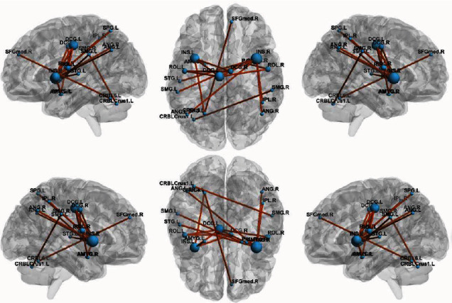
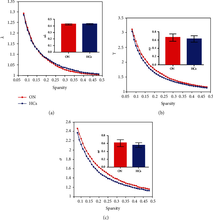
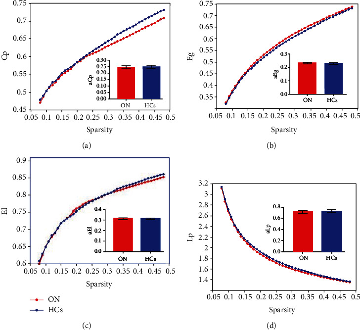
In the sparsity range from 0.08 to 0.48, both groups exhibited small-world attributes. Compared to the control group, global network efficiency, normalized clustering coefficient, and small-world value were higher whereas the clustering coefficient value was lower in ON patients. There were no differences in characteristic path length, local network efficiency, and normalized characteristic path length between groups. In addition, ON patients had lower brain functional connectivity strength among the rolandic operculum, medial superior frontal gyrus, insula, median cingulate and paracingulate gyri, amygdala, superior parietal gyrus, inferior parietal gyrus, supramarginal gyrus, angular gyrus, lenticular nucleus, pallidum, superior temporal gyrus, and cerebellum compared to the control group ( < 0.05).

CONCLUSION

Patients with ON show typical "small world" topology that differed from that detected in HC brain networks. The brain network in ON has a small-world attribute but shows reduced and abnormal connectivity compared to normal subjects and likely causes symptoms of cognitive impairment.

摘要

目的

本研究通过静息态功能磁共振成像(rs-fMRI)和基于图论的方法,研究视神经炎(ON)患者小世界拓扑结构和脑功能连接的变化。

方法

共纳入 21 例 ON 患者(8 名男性,13 名女性)和 21 名匹配的健康对照者(8 名男性,13 名女性),并进行 rs-fMRI 检查。对数据进行预处理,将大脑分为 116 个感兴趣区。从成对脑区间相关系数计算小世界网络参数和曲线下面积(AUC)。采用独立样本 t 检验比较两组脑网络参数 AUC 的差异,采用基于网络的统计分析评估 ON 患者与对照组之间脑连接强度的变化。

结果

在稀疏度范围为 0.08 至 0.48 时,两组均表现出小世界属性。与对照组相比,ON 患者的全局网络效率、标准化聚类系数和小世界值较高,而聚类系数值较低。组间特征路径长度、局部网络效率和标准化特征路径长度无差异。此外,与对照组相比,ON 患者大脑中 Rolandic 盖、内侧额上回、岛叶、中央扣带回和旁扣带回、杏仁核、顶上小叶、顶下小叶、缘上回、角回、豆状核、苍白球、颞上回和小脑的脑功能连接强度较低(<0.05)。

结论

ON 患者表现出典型的“小世界”拓扑结构,与 HC 脑网络中检测到的不同。ON 患者的脑网络具有小世界属性,但与正常受试者相比,连接减少且异常,可能导致认知障碍症状。

https://cdn.ncbi.nlm.nih.gov/pmc/blobs/b6dc/8219459/62f11f059c2b/DM2021-9948751.004.jpg
https://cdn.ncbi.nlm.nih.gov/pmc/blobs/b6dc/8219459/1363594d9eb2/DM2021-9948751.001.jpg
https://cdn.ncbi.nlm.nih.gov/pmc/blobs/b6dc/8219459/eb073592e874/DM2021-9948751.002.jpg
https://cdn.ncbi.nlm.nih.gov/pmc/blobs/b6dc/8219459/b42eb5f13bcc/DM2021-9948751.003.jpg
https://cdn.ncbi.nlm.nih.gov/pmc/blobs/b6dc/8219459/62f11f059c2b/DM2021-9948751.004.jpg

相似文献

1
Altered Small-World Functional Network Topology in Patients with Optic Neuritis: A Resting-State fMRI Study.视神经炎患者的小世界功能网络拓扑结构改变:一项静息态 fMRI 研究。
Dis Markers. 2021 Jun 14;2021:9948751. doi: 10.1155/2021/9948751. eCollection 2021.
2
Acute Changes in the Resting Brain Networks in Concussion Patients: Small-World Topology Perspective.脑震荡患者静息态脑网络的急性变化:小世界拓扑结构视角。
J Integr Neurosci. 2024 Jan 16;23(1):12. doi: 10.31083/j.jin2301012.
3
[Analysis of the Characteristics of Infantile Small World Neural Network Node Properties Correlated with the Influencing Factors].[与影响因素相关的小儿小世界神经网络节点特性分析]
Sheng Wu Yi Xue Gong Cheng Xue Za Zhi. 2016 Oct;33(5):931-8.
4
Brain functional connectivity during storage based on resting state functional magnetic resonance imaging with synchronous urodynamic testing in healthy volunteers.基于静息态功能磁共振成像和健康志愿者同步尿动力学测试的储存期脑功能连接。
Brain Imaging Behav. 2021 Jun;15(3):1676-1684. doi: 10.1007/s11682-020-00362-y.
5
A Graph Theory Study of Resting-State Functional MRI Connectivity in Children With Carbon Monoxide Poisoning.一氧化碳中毒儿童静息态功能磁共振成像连接性的图论研究
J Magn Reson Imaging. 2023 Nov;58(5):1452-1459. doi: 10.1002/jmri.28706. Epub 2023 Mar 30.
6
[Changes of brain structural network properties in patients with rapid eye movement sleep behavior disorder].[快速眼动睡眠行为障碍患者脑结构网络属性的变化]
Nan Fang Yi Ke Da Xue Xue Bao. 2020 Jan 30;40(1):125-130. doi: 10.12122/j.issn.1673-4254.2020.01.20.
7
Reduced resting-state brain functional network connectivity and poor regional homogeneity in patients with CADASIL.CADASIL 患者静息态脑功能网络连接减少和局部一致性降低。
J Headache Pain. 2019 Nov 11;20(1):103. doi: 10.1186/s10194-019-1052-6.
8
Graph theory analysis of resting-state functional magnetic resonance imaging in essential tremor.静息态功能磁共振成像在特发性震颤中的图论分析。
Hum Brain Mapp. 2019 Nov 1;40(16):4686-4702. doi: 10.1002/hbm.24730. Epub 2019 Jul 22.
9
Disrupted brain network topology in pediatric posttraumatic stress disorder: A resting-state fMRI study.儿童创伤后应激障碍中大脑网络拓扑结构的破坏:一项静息态功能磁共振成像研究。
Hum Brain Mapp. 2015 Sep;36(9):3677-86. doi: 10.1002/hbm.22871. Epub 2015 Jun 19.
10
Alternations of interhemispheric functional connectivity in patients with optic neuritis using voxel-mirrored homotopic connectivity: A resting state fMRI study.基于体素镜像同伦连接的静息态 fMRI 研究:视神经炎患者大脑两半球间功能连接的改变。
Brain Imaging Behav. 2023 Feb;17(1):1-10. doi: 10.1007/s11682-022-00719-5. Epub 2022 Nov 28.

引用本文的文献

1
Abnormal topological properties and functional-structural coupling of large-scale brain networks in primary angle-closure glaucoma.原发性闭角型青光眼患者大脑大规模网络的异常拓扑特性及功能-结构耦合
Quant Imaging Med Surg. 2025 Jul 1;15(7):6053-6067. doi: 10.21037/qims-24-1947. Epub 2025 Jun 30.
2
Abnormal intrinsic brain functional network dynamics in patients with retinal detachment based on graph theory and machine learning.基于图论和机器学习的视网膜脱离患者大脑内在功能网络动力学异常
Heliyon. 2024 Nov 2;10(23):e37890. doi: 10.1016/j.heliyon.2024.e37890. eCollection 2024 Dec 15.
3
Frequency-specific segregation and integration of human cerebral cortex: An intrinsic functional atlas.

本文引用的文献

1
Bilateral retinal pathology following a first-ever clinical episode of autoimmune optic neuritis.首例自身免疫性视神经炎临床发作后的双侧视网膜病变。
Neurol Neuroimmunol Neuroinflamm. 2020 Jan 22;7(2). doi: 10.1212/NXI.0000000000000671. Print 2020 Mar 5.
2
Activation of EphB2 in the basolateral amygdala promotes stress vulnerability of mice by increasing NMDA-dependent synaptic function.外侧杏仁核中 EphB2 的激活通过增加 NMDA 依赖性突触功能促进了小鼠的应激易感性。
Neuropharmacology. 2020 May 1;167:107934. doi: 10.1016/j.neuropharm.2019.107934. Epub 2020 Jan 9.
3
Brain Atrophy in Relapsing Optic Neuritis Is Associated With Crion Phenotype.
人类大脑皮层的频率特异性分离与整合:一个内在功能图谱。
iScience. 2024 Feb 19;27(3):109206. doi: 10.1016/j.isci.2024.109206. eCollection 2024 Mar 15.
4
Topological properties and connectivity patterns in brain networks of patients with refractory epilepsy combined with intracranial electrical stimulation.难治性癫痫患者脑网络的拓扑性质和连接模式与颅内电刺激的相关性
Front Neurosci. 2023 Nov 23;17:1282232. doi: 10.3389/fnins.2023.1282232. eCollection 2023.
5
[Changes of brain network in patients with insomnia following radiotherapy for cervical cancer and their correlation with IT-TMS treatment efficacy: a graph-theory analysis].宫颈癌放疗后失眠患者脑网络变化及其与重复经颅磁刺激治疗疗效的相关性:基于图论的分析
Nan Fang Yi Ke Da Xue Xue Bao. 2023 Sep 20;43(9):1629-1635. doi: 10.12122/j.issn.1673-4254.2023.09.22.
6
Graph-Theory-Based Degree Centrality Combined with Machine Learning Algorithms Can Predict Response to Treatment with Antipsychotic Medications in Patients with First-Episode Schizophrenia.基于图论的度数中心度结合机器学习算法可预测首发精神分裂症患者对抗精神病药物治疗的反应。
Dis Markers. 2022 Oct 13;2022:1853002. doi: 10.1155/2022/1853002. eCollection 2022.
7
Frequency-Specific Analysis of the Dynamic Reconfiguration of the Brain in Patients with Schizophrenia.精神分裂症患者大脑动态重构的频率特异性分析
Brain Sci. 2022 Jun 1;12(6):727. doi: 10.3390/brainsci12060727.
8
Altered Brain Activity in Patients With Comitant Strabismus Detected by Analysis of the Fractional Amplitude of Low-Frequency Fluctuation: A Resting-State Functional MRI Study.基于低频波动分数振幅分析检测的伴有斜视患者的脑活动改变:一项静息态功能磁共振成像研究
Front Hum Neurosci. 2022 Apr 8;16:874703. doi: 10.3389/fnhum.2022.874703. eCollection 2022.
9
Brain Network Topology and Structural-Functional Connectivity Coupling Mediate the Association Between Gut Microbiota and Cognition.脑网络拓扑结构与结构-功能连接耦合介导肠道微生物群与认知之间的关联。
Front Neurosci. 2022 Mar 29;16:814477. doi: 10.3389/fnins.2022.814477. eCollection 2022.
10
Improved Functional Organization in Patients With Primary Insomnia After Individually-Targeted Transcranial Magnetic Stimulation.个体化靶向经颅磁刺激后原发性失眠患者功能组织的改善
Front Neurosci. 2022 Mar 10;16:859440. doi: 10.3389/fnins.2022.859440. eCollection 2022.
复发性视神经炎中的脑萎缩与Crion表型相关。
Front Neurol. 2019 Nov 1;10:1157. doi: 10.3389/fneur.2019.01157. eCollection 2019.
4
Small-world properties of the whole-brain functional networks in patients with obstructive sleep apnea-hypopnea syndrome.阻塞性睡眠呼吸暂停低通气综合征患者全脑功能网络的小世界属性。
Sleep Med. 2019 Oct;62:53-58. doi: 10.1016/j.sleep.2018.08.037. Epub 2018 Dec 12.
5
Effect of second-generation antipsychotics on brain network topology in first-episode schizophrenia: A longitudinal rs-fMRI study.第二代抗精神病药对首发精神分裂症脑网络拓扑结构的影响:一项纵向 rs-fMRI 研究。
Schizophr Res. 2019 Jun;208:160-166. doi: 10.1016/j.schres.2019.03.015. Epub 2019 Apr 7.
6
Increased Small-World Network Topology Following Deployment-Acquired Traumatic Brain Injury Associated with the Development of Post-Traumatic Stress Disorder.创伤性脑损伤后创伤后应激障碍相关的与部署获得性创伤性脑损伤后的小世界网络拓扑结构增加。
Brain Connect. 2018 May;8(4):205-211. doi: 10.1089/brain.2017.0556. Epub 2018 Apr 20.
7
Gemcitabine directly inhibits effector CD4 T cell activation and prevents experimental autoimmune encephalomyelitis.吉西他滨直接抑制效应性 CD4 T 细胞活化,防止实验性自身免疫性脑脊髓炎。
J Neuroimmunol. 2018 Mar 15;316:7-16. doi: 10.1016/j.jneuroim.2017.12.002. Epub 2017 Dec 8.
8
Disrupted small-world brain functional network topology in male patients with severe obstructive sleep apnea revealed by resting-state fMRI.静息态功能磁共振成像揭示重度阻塞性睡眠呼吸暂停男性患者小世界脑功能网络拓扑结构破坏
Neuropsychiatr Dis Treat. 2017 Jun 8;13:1471-1482. doi: 10.2147/NDT.S135426. eCollection 2017.
9
Correlation between standardized assessment of concussion scores and small-world brain network in mild traumatic brain injury.轻度创伤性脑损伤中脑震荡评分的标准化评估与小世界脑网络之间的相关性。
J Clin Neurosci. 2017 Oct;44:114-121. doi: 10.1016/j.jocn.2017.05.010. Epub 2017 Jun 8.
10
Small world brain network characteristics during EEG Holter recording of a stroke event.中风事件脑电图动态记录期间的小世界脑网络特征
Clin Neurophysiol. 2017 Jan;128(1):1-3. doi: 10.1016/j.clinph.2016.10.090. Epub 2016 Nov 3.