Friedrich Miescher Institute for Biomedical Research, Basel, Switzerland.
Department of Pharmacology and Toxicology, Institute of Pharmacy and CMBI, University of Innsbruck, Innsbruck, Austria.
Nat Commun. 2021 Jul 6;12(1):4156. doi: 10.1038/s41467-021-24068-x.
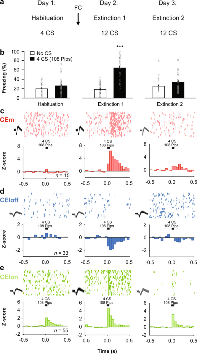
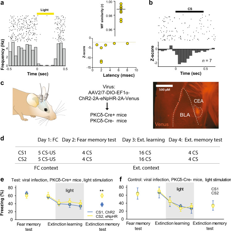
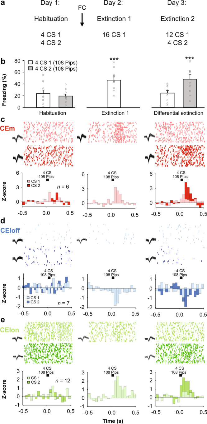
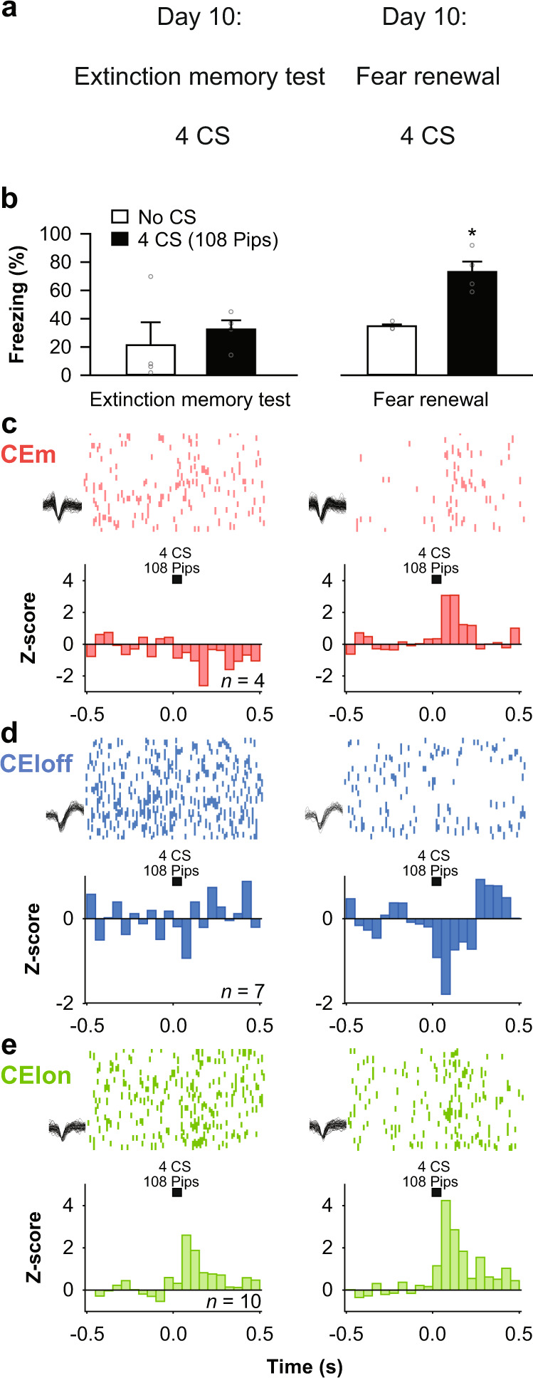
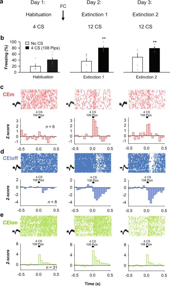
Fear extinction is an adaptive process whereby defensive responses are attenuated following repeated experience of prior fear-related stimuli without harm. The formation of extinction memories involves interactions between various corticolimbic structures, resulting in reduced central amygdala (CEA) output. Recent studies show, however, the CEA is not merely an output relay of fear responses but contains multiple neuronal subpopulations that interact to calibrate levels of fear responding. Here, by integrating behavioural, in vivo electrophysiological, anatomical and optogenetic approaches in mice we demonstrate that fear extinction produces reversible, stimulus- and context-specific changes in neuronal responses to conditioned stimuli in functionally and genetically defined cell types in the lateral (CEl) and medial (CEm) CEA. Moreover, we show these alterations are absent when extinction is deficient and that selective silencing of protein kinase C delta-expressing (PKCδ) CEl neurons impairs fear extinction. Our findings identify CEA inhibitory microcircuits that act as critical elements within the brain networks mediating fear extinction.
恐惧消退是一种适应性过程,即在没有伤害的情况下,通过反复经历先前与恐惧相关的刺激,防御反应会减弱。消退记忆的形成涉及各种皮质边缘结构之间的相互作用,导致中央杏仁核(CEA)输出减少。然而,最近的研究表明,CEA 不仅仅是恐惧反应的输出中继,它还包含多个神经元亚群,这些亚群相互作用以调节恐惧反应的水平。在这里,我们通过整合行为学、在体电生理学、解剖学和光遗传学方法,在小鼠中证明,恐惧消退会导致对条件刺激的神经元反应产生可逆的、刺激特异性和情境特异性变化,这些变化存在于功能和基因定义的外侧(CEl)和内侧(CEm)CEA 中的细胞类型中。此外,我们还表明,当消退不足时,这些改变是不存在的,并且选择性沉默表达蛋白激酶 C 三角洲(PKCδ)的 CEl 神经元会损害恐惧消退。我们的发现确定了 CEA 抑制性微电路,它们作为介导恐惧消退的大脑网络中的关键元素发挥作用。