College of Engineering, Swansea University, Swansea, UK.
Department of Veterinary Medicine, Cambridge University, Cambridge, UK.
Arch Toxicol. 2021 Sep;95(9):3101-3115. doi: 10.1007/s00204-021-03113-0. Epub 2021 Jul 10.
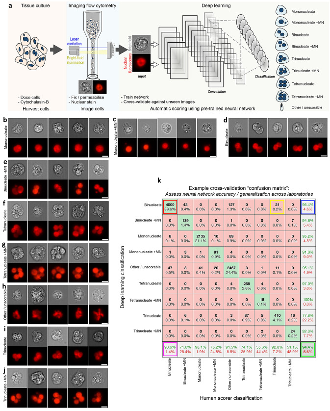
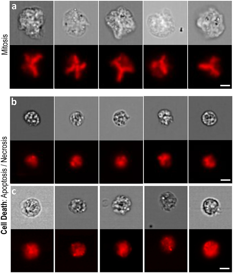
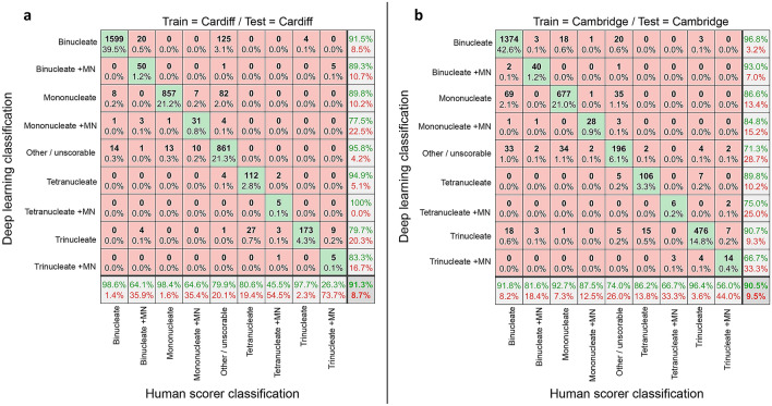
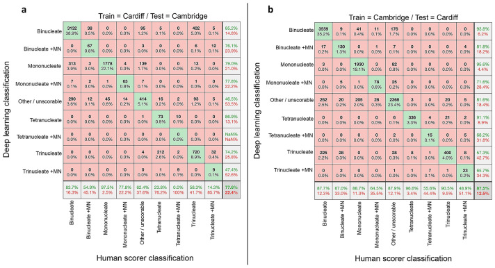
The in vitro micronucleus assay is a globally significant method for DNA damage quantification used for regulatory compound safety testing in addition to inter-individual monitoring of environmental, lifestyle and occupational factors. However, it relies on time-consuming and user-subjective manual scoring. Here we show that imaging flow cytometry and deep learning image classification represents a capable platform for automated, inter-laboratory operation. Images were captured for the cytokinesis-block micronucleus (CBMN) assay across three laboratories using methyl methanesulphonate (1.25-5.0 μg/mL) and/or carbendazim (0.8-1.6 μg/mL) exposures to TK6 cells. Human-scored image sets were assembled and used to train and test the classification abilities of the "DeepFlow" neural network in both intra- and inter-laboratory contexts. Harnessing image diversity across laboratories yielded a network able to score unseen data from an entirely new laboratory without any user configuration. Image classification accuracies of 98%, 95%, 82% and 85% were achieved for 'mononucleates', 'binucleates', 'mononucleates with MN' and 'binucleates with MN', respectively. Successful classifications of 'trinucleates' (90%) and 'tetranucleates' (88%) in addition to 'other or unscorable' phenotypes (96%) were also achieved. Attempts to classify extremely rare, tri- and tetranucleated cells with micronuclei into their own categories were less successful (≤ 57%). Benchmark dose analyses of human or automatically scored micronucleus frequency data yielded quantitation of the same equipotent concentration regardless of scoring method. We conclude that this automated approach offers significant potential to broaden the practical utility of the CBMN method across industry, research and clinical domains. We share our strategy using openly-accessible frameworks.
体外微核试验是一种用于监管化合物安全性测试的重要的全球方法,除此之外,还可以用于个体间的环境、生活方式和职业因素的监测。然而,它依赖于耗时且需要用户主观判断的手动评分。在这里,我们展示了成像流式细胞术和深度学习图像分类代表了一种用于自动化、实验室间操作的可行平台。使用甲基甲烷磺酸盐(1.25-5.0μg/mL)和/或多菌灵(0.8-1.6μg/mL)暴露于 TK6 细胞,在三个实验室中捕获了胞质分裂阻断微核(CBMN)试验的图像。为了在实验室内部和实验室之间训练和测试“DeepFlow”神经网络的分类能力,我们收集了人工评分的图像集。利用实验室之间的图像多样性,开发出了一种能够对全新实验室的未见过数据进行评分的网络,而无需任何用户配置。对于“单核细胞”、“双核细胞”、“单核细胞带有微核”和“双核细胞带有微核”,图像分类准确率分别达到了 98%、95%、82%和 85%。此外,还成功地对“三核细胞”(90%)和“四核细胞”(88%)以及“其他或不可评分”表型(96%)进行了分类。对带有微核的非常罕见的三核和四核细胞进行分类的尝试则不太成功(≤57%)。对人工或自动评分的微核频率数据进行基准剂量分析,得出了无论评分方法如何,相同效等量浓度的定量结果。我们得出结论,这种自动化方法具有广泛的潜力,可以拓宽 CBMN 方法在工业、研究和临床领域的实际应用。我们共享了使用开放获取框架的策略。