Division of Radiology, Geneva University Hospitals, Ch. du Pont-Bochet 3, 1226, Thonex, Switzerland.
Division of Radiology, University of Geneva, Rue Gabrielle-Perret-Gentil 4, 1205, Geneva, Switzerland.
BMC Med Imaging. 2021 Jul 12;21(1):110. doi: 10.1186/s12880-021-00641-0.
For the treatment of radicular pain, nerve root infiltrations can be performed under MRI guidance in select, typically younger, patients where repeated CT exams are not desirable due to associated radiation risk, or potential allergic reactions to iodinated contrast medium.
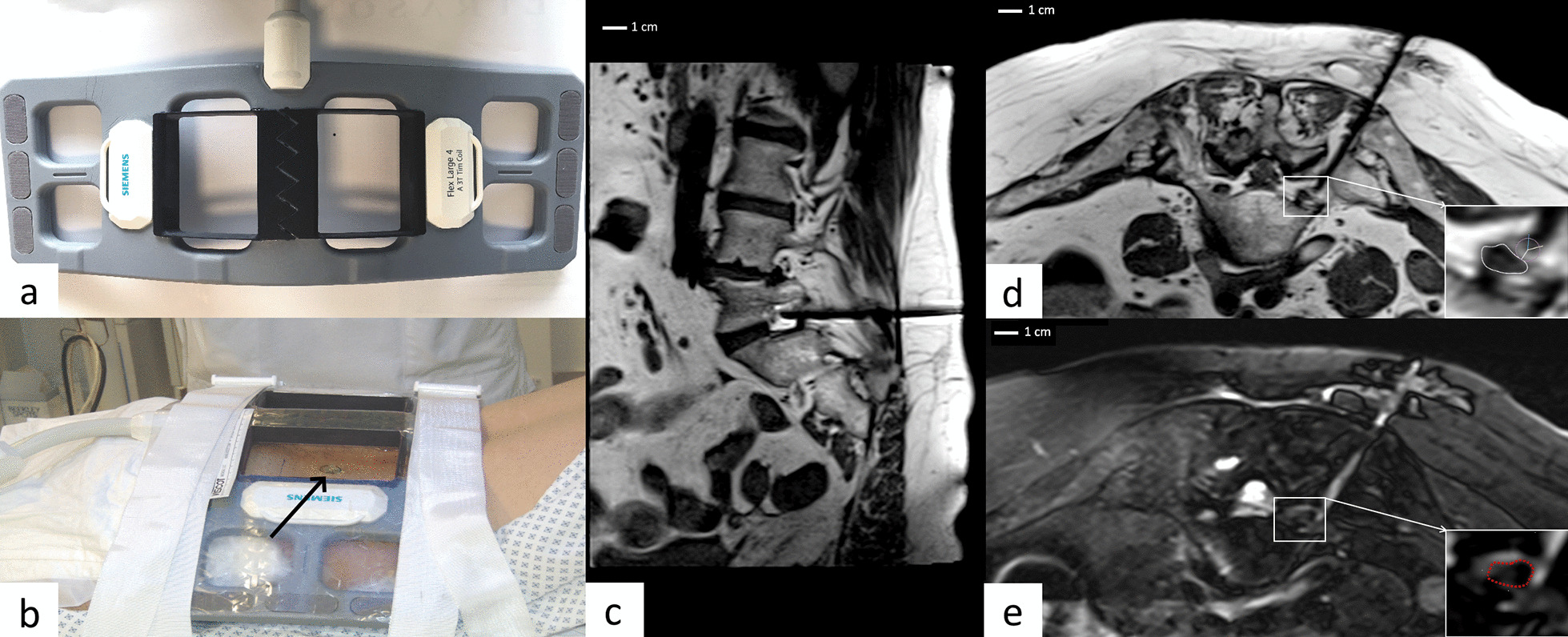
Fifteen 3 T MRI-guided nerve root infiltrations were performed in 12 patients with a dedicated surface coil combined with the standard spine coil, using a breathhold PD sequence. The needle artifact on the MR images and the distance between the needle tip and the infiltrated nerve root were measured.
The distance between the needle tip and the nerve root was 2.1 ± 1.4 mm. The visual artifact width, perpendicular to the needle long axis, was 2.1 ± 0.7 mm. No adverse events were reported.
This technical note describes the optimization of the procedure in a 3 T magnetic field, including reported procedure time and an assessment of targeting precision.
对于神经根痛的治疗,可以在特定的、通常是年轻的患者中,在 MRI 引导下进行神经根浸润,这些患者由于与辐射风险相关,或对碘造影剂有潜在的过敏反应,不适合进行多次 CT 检查。
在 12 名患者中进行了 15 次 3T MRI 引导的神经根浸润,使用专用表面线圈结合标准脊柱线圈,采用屏气 PD 序列。测量了 MR 图像上的针迹伪影和针尖与浸润神经根之间的距离。
针尖与神经根之间的距离为 2.1±1.4mm。垂直于针长轴的视觉伪影宽度为 2.1±0.7mm。未报告不良事件。
本技术说明描述了在 3T 磁场中优化该程序,包括报告的程序时间和靶向精度评估。