Vilnius University, Vilnius, Lithuania.
Stanford Nano Shared Facilities, Stanford University, Stanford, USA.
Sci Rep. 2021 Jul 20;11(1):14810. doi: 10.1038/s41598-021-94303-4.
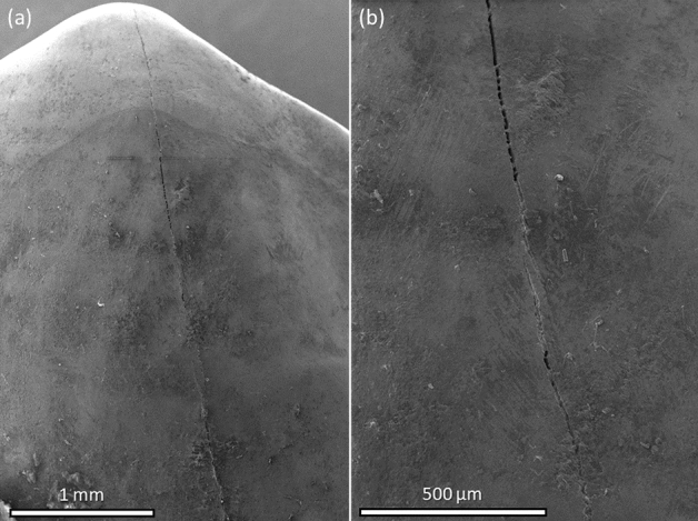
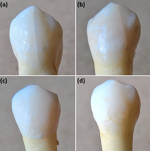
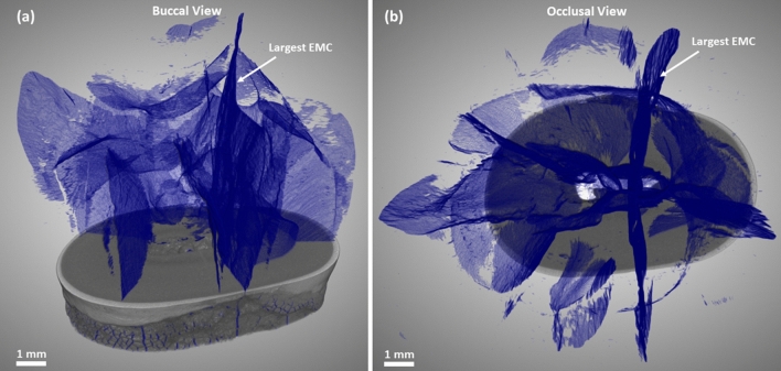
Although the topic of tooth fractures has been extensively analyzed in the dental literature, there is still insufficient information about the potential effect of enamel microcracks (EMCs) on the underlying tooth structures. For a precise examination of the extent of the damage to the tooth structure in the area of EMCs, it is necessary to carry out their volumetric [(three-dimensional (3D)] evaluation. The aim of this study was to validate an X-ray micro-computed tomography ([Formula: see text]CT) as a technique suitable for 3D non-destructive visualization and qualitative analysis of teeth EMCs of different severity. Extracted human maxillary premolars were examined using a [Formula: see text]CT instrument ZEISS Xradia 520 Versa. In order to separate crack, dentin, and enamel volumes a Deep Learning (DL) algorithm, part of the Dragonfly's segmentation toolkit, was utilized. For segmentation needs we implemented Dragonfly's pre-built UNet neural network. The scanning technique which was used made it possible to recognize and detect not only EMCs that are visible on the outer surface but also those that are buried deep inside the tooth. The 3D visualization, combined with DL assisted segmentation, enabled the evaluation of the dynamics of an EMC and precise examination of its position with respect to the dentin-enamel junction.
虽然牙齿折裂这一主题在牙科文献中已经被广泛分析,但关于牙釉质微裂(EMC)对下方牙齿结构潜在影响的信息仍然不足。为了精确检查 EMC 区域内牙齿结构的损伤程度,有必要对其进行体积[(三维(3D)]评估。本研究的目的是验证 X 射线微计算机断层扫描 ([Formula: see text]CT) 作为一种适合 EMC 不同严重程度的 3D 无损可视化和定性分析的技术。使用蔡司 Xradia 520 Versa 型 [Formula: see text]CT 仪器对提取的上颌前磨牙进行了检查。为了分离裂纹、牙本质和牙釉质体积,使用了深度学习(DL)算法,该算法是 Dragonfly 分割工具包的一部分。为了满足分割需求,我们实现了 Dragonfly 预先构建的 UNet 神经网络。所使用的扫描技术不仅可以识别和检测外表面可见的 EMC,还可以检测深埋在牙齿内部的 EMC。3D 可视化结合 DL 辅助分割,实现了 EMC 动力学的评估,并可以精确检查其相对于牙本质-釉质交界处的位置。