Department of Plastic Surgery, Hand and Burn Surgery, Burn Center, Rhein Maas Klinikum, 52146 Wuerselen, Germany.
Plastic Surgery and Burn Unit, Fakeeh Care & Fakeeh College of Medical Sciences, P.O. Box 2537, Jeddah 21461, Saudi Arabia.
Med Sci (Basel). 2021 Jul 16;9(3):51. doi: 10.3390/medsci9030051.
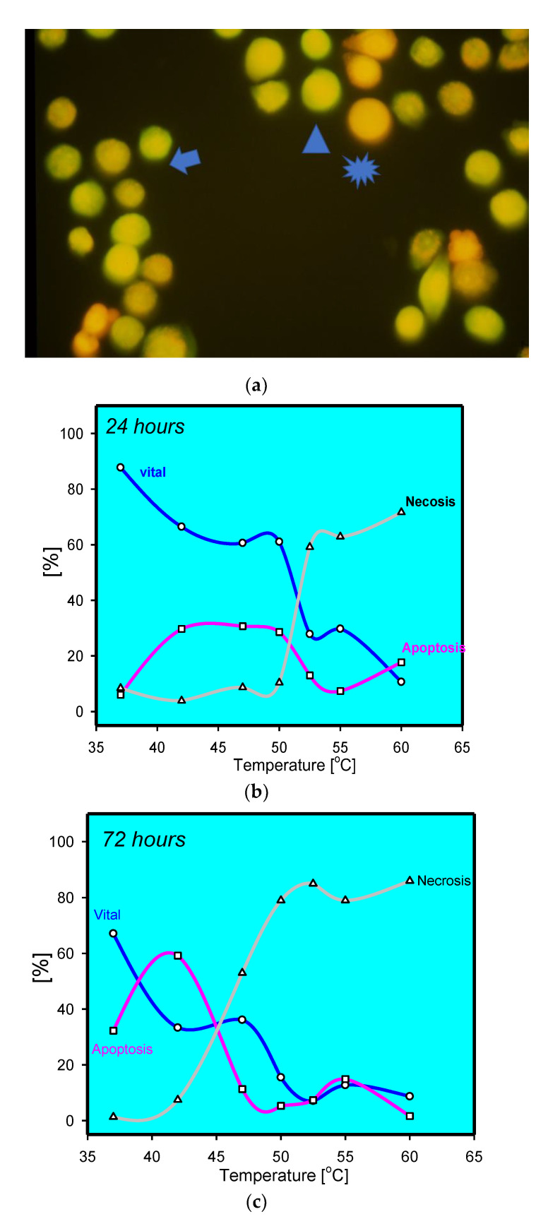
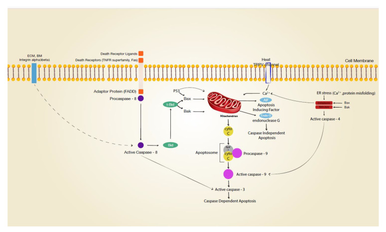
Cutaneous burn injury is associated with epidermal loss in the zone of coagulation zone and delayed tissue loss in the zone of stasis. Thus, thermal stress can trigger both necrosis and regulated cell death (RCD) or apoptosis. Experimental in vitro and in vivo work has clearly demonstrated apoptotic events of thermally injured keratinocytes that are accompanied by morphological and biochemical markers of regulated cell death. However, in vivo data for the different pathways of regulated cell death are sparse. In vitro experiments with heat-stressed human keratinocytes have demonstrated death receptor involvement (extrinsic apoptosis), calcium influx, and disruption of mitochondrial membrane potential (intrinsic apoptosis) in regulated cell death. In addition, caspase-independent pathways have been suggested in regulated cell death. Keratinocyte heat stress leads to reduced proliferation, possibly as a result of reduced keratinocyte adhesion (anoikis) or oncogene involvement. Understanding the underlying mechanisms of RCD and the skin's responses to thermal stress may lead to improved strategies for treating cutaneous burn trauma.
皮肤烧伤损伤与凝固区的表皮丧失以及淤滞区的延迟组织丧失有关。因此,热应激可引发细胞坏死和细胞程序性死亡(RCD)或细胞凋亡。实验室内和体内研究工作清楚地表明,热烧伤角质形成细胞存在细胞程序性死亡的事件,同时伴有形态和生化标记物的变化。然而,关于不同途径的细胞程序性死亡的体内数据很少。体外实验表明,热应激的人角质形成细胞存在死亡受体的参与(外在细胞凋亡)、钙内流和线粒体膜电位的破坏(内在细胞凋亡)。此外,还提出了无半胱氨酸天冬氨酸蛋白酶的途径参与细胞程序性死亡。角质形成细胞的热应激导致增殖减少,可能是由于角质形成细胞黏附(失巢凋亡)减少或癌基因的参与。了解细胞程序性死亡的潜在机制以及皮肤对热应激的反应可能会导致治疗皮肤烧伤创伤的策略得到改善。