Department of Internal Medicine, Division of Rheumatology, Allergy, and Clinical Immunology, University of California, Davis, CA, USA.
MIND Institute, University of California, Davis, CA, USA.
Mol Psychiatry. 2021 Dec;26(12):7530-7537. doi: 10.1038/s41380-021-01215-w. Epub 2021 Jul 21.
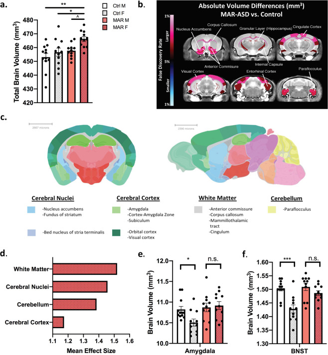
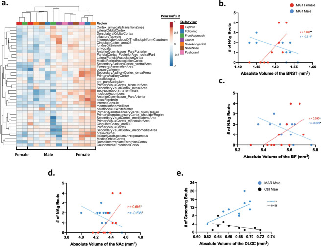
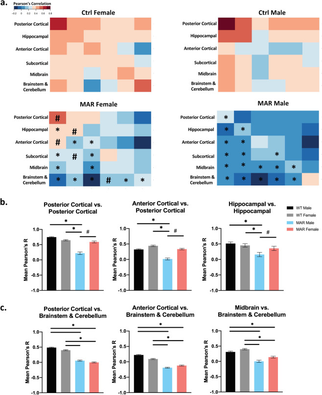
Immunoglobulin G (IgG) autoantibodies reactive to fetal brain proteins in mothers of children with ASD have been described by several groups. To understand their pathologic significance, we developed a mouse model of maternal autoantibody related ASD (MAR-ASD) utilizing the peptide epitopes from human autoantibody reactivity patterns. Male and female offspring prenatally exposed to the salient maternal autoantibodies displayed robust deficits in social interactions and increased repetitive self-grooming behaviors as juveniles and adults. In the present study, neuroanatomical differences in adult MAR-ASD and control offspring were assessed via high-resolution ex vivo magnetic resonance imaging (MRI) at 6 months of age. Of interest, MAR-ASD mice displayed significantly larger total brain volume and of the 159 regions examined, 31 were found to differ significantly in absolute volume (mm) at an FDR of <5%. Specifically, the absolute volumes of several white matter tracts, cortical regions, and basal nuclei structures were significantly increased in MAR-ASD animals. These phenomena were largely driven by female MAR-ASD offspring, as no significant differences were seen with either absolute or relative regional volume in male MAR-ASD mice. However, structural covariance analysis suggests network-level desynchronization in brain volume in both male and female MAR-ASD mice. Additionally, preliminary correlational analysis with behavioral data relates that volumetric increases in numerous brain regions of MAR-ASD mice were correlated with social interaction and repetitive self-grooming behaviors in a sex-specific manner. These results demonstrate significant sex-specific effects in brain size, regional relationships, and behavior for offspring prenatally exposed to MAR-ASD autoantibodies relative to controls.
免疫球蛋白 G(IgG)自身抗体与自闭症谱系障碍(ASD)儿童的母亲的胎儿脑蛋白反应已被几个研究小组描述。为了了解其病理意义,我们利用人类自身抗体反应模式的肽表位,开发了一种与母体自身抗体相关的自闭症谱系障碍(MAR-ASD)的小鼠模型。产前暴露于显著母体自身抗体的雄性和雌性后代在青少年和成年时表现出强烈的社交互动缺陷和增加的重复自我修饰行为。在本研究中,通过 6 个月龄时的高分辨率离体磁共振成像(MRI)评估成年 MAR-ASD 和对照后代的神经解剖差异。有趣的是,MAR-ASD 小鼠的总脑体积明显增大,在检查的 159 个区域中,有 31 个区域的绝对体积(mm)在 FDR <5%时存在显著差异。具体而言,MAR-ASD 动物的几个白质束、皮质区域和基底核结构的绝对体积显著增加。这些现象主要是由雌性 MAR-ASD 后代驱动的,因为在雄性 MAR-ASD 小鼠中,无论是绝对还是相对区域体积都没有观察到显著差异。然而,结构协方差分析表明,雄性和雌性 MAR-ASD 小鼠的大脑体积存在网络级别的去同步化。此外,与行为数据的初步相关分析表明,MAR-ASD 小鼠许多脑区的体积增加与社交互动和重复自我修饰行为呈性别特异性相关。这些结果表明,与对照组相比,产前暴露于 MAR-ASD 自身抗体的后代的大脑大小、区域关系和行为存在显著的性别特异性影响。