• 文献检索
  • 文档翻译
  • 深度研究
  • 学术资讯
  • Suppr Zotero 插件Zotero 插件
  • 邀请有礼
  • 套餐&价格
  • 历史记录
应用&插件
Suppr Zotero 插件Zotero 插件浏览器插件Mac 客户端Windows 客户端微信小程序
定价
高级版会员购买积分包购买API积分包
服务
文献检索文档翻译深度研究API 文档MCP 服务
关于我们
关于 Suppr公司介绍联系我们用户协议隐私条款
关注我们

Suppr 超能文献

核心技术专利:CN118964589B侵权必究
粤ICP备2023148730 号-1Suppr @ 2026

文献检索

告别复杂PubMed语法,用中文像聊天一样搜索,搜遍4000万医学文献。AI智能推荐,让科研检索更轻松。

立即免费搜索

文件翻译

保留排版,准确专业,支持PDF/Word/PPT等文件格式,支持 12+语言互译。

免费翻译文档

深度研究

AI帮你快速写综述,25分钟生成高质量综述,智能提取关键信息,辅助科研写作。

立即免费体验

晚期糖尿病肾病中RhoGAP Myosin9A的S-亚硝基化发生改变。

S-Nitrosylation of RhoGAP Myosin9A Is Altered in Advanced Diabetic Kidney Disease.

作者信息

Li Qi, Veron Delma, Tufro Alda

机构信息

Department of Pediatrics/Nephrology, New Haven, CT, United States.

Department of Cell and Molecular Physiology, Yale School of Medicine, New Haven, CT, United States.

出版信息

Front Med (Lausanne). 2021 Jul 14;8:679518. doi: 10.3389/fmed.2021.679518. eCollection 2021.

DOI:10.3389/fmed.2021.679518
PMID:34336885
原文链接:https://pmc.ncbi.nlm.nih.gov/articles/PMC8316719/
Abstract

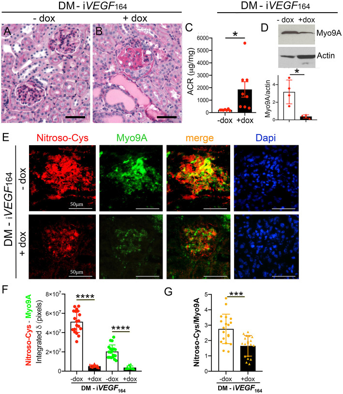
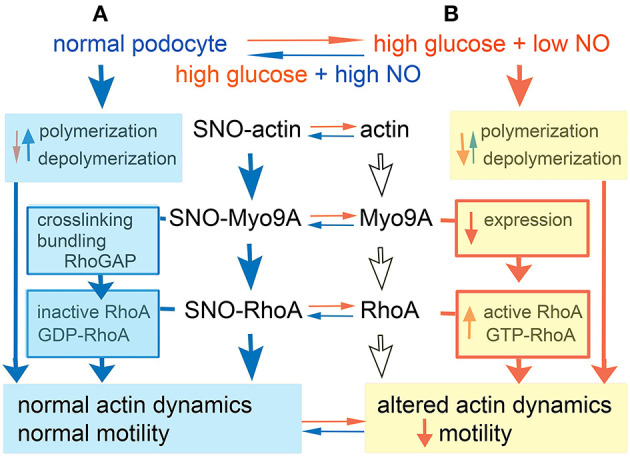
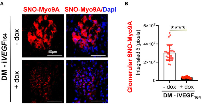
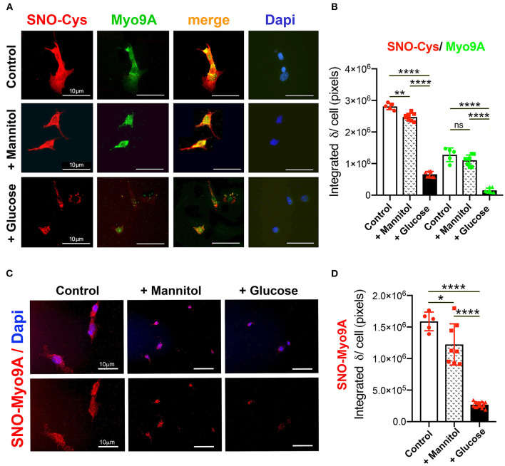
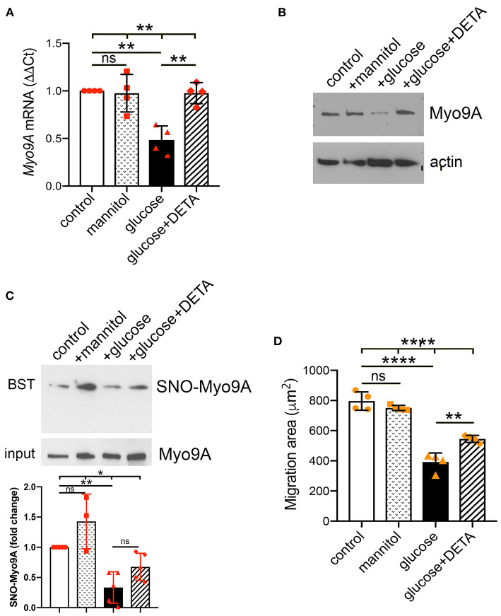
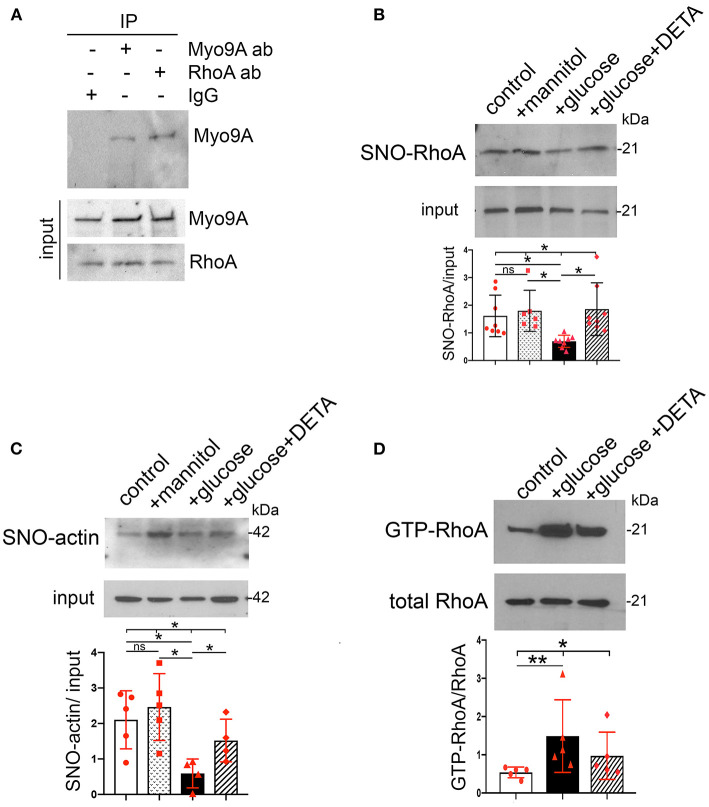
The molecular pathogenesis of diabetic kidney disease progression is complex and remains unresolved. Rho-GAP was recently identified as a novel podocyte protein and a candidate gene for monogenic FSGS. Myo9A involvement in diabetic kidney disease has been suggested. Here, we examined the effect of diabetic milieu on Myo9A expression and . We determined that Myo9A undergoes S-nitrosylation, a post-translational modification dependent on nitric oxide (NO) availability. Diabetic mice with nodular glomerulosclerosis and severe proteinuria associated with doxycycline-induced, podocyte-specific gain-of-function showed markedly decreased glomerular Myo9A expression and S-nitrosylation, as compared to uninduced diabetic mice. Immortalized mouse podocytes exposed to high glucose revealed decreased expression, assessed by qPCR, immunoblot and immunocytochemistry, and reduced Myo9A S-nitrosylation (SNO-Myo9A), assessed by proximity link assay and biotin switch test, functionally resulting in abnormal podocyte migration. These defects were abrogated by exposure to a NO donor and were not due to hyperosmolarity. Our data demonstrate that high-glucose induced decrease of both expression and SNO-Myo9A is regulated by NO availability. We detected S-nitrosylation of Myo9A interacting proteins RhoA and actin, which was also altered by high glucose and NO dependent. RhoA activity inversely related to SNO-RhoA. Collectively, data suggest that dysregulation of SNO-Myo9A, SNO-RhoA and SNO-actin may contribute to the pathogenesis of advanced diabetic kidney disease and may be amenable to therapeutic targeting.

摘要

糖尿病肾病进展的分子发病机制复杂,尚未得到解决。Rho-GAP最近被鉴定为一种新型足细胞蛋白和单基因FSGS的候选基因。已有研究表明Myo9A参与糖尿病肾病。在此,我们研究了糖尿病环境对Myo9A表达的影响。我们确定Myo9A会发生S-亚硝基化,这是一种依赖于一氧化氮(NO)可用性的翻译后修饰。与未诱导的糖尿病小鼠相比,在强力霉素诱导的足细胞特异性功能获得的情况下,出现结节性肾小球硬化和严重蛋白尿的糖尿病小鼠肾小球Myo9A表达和S-亚硝基化明显降低。通过qPCR、免疫印迹和免疫细胞化学评估,暴露于高糖环境的永生化小鼠足细胞Myo9A表达降低,通过邻近连接分析和生物素开关试验评估,Myo9A的S-亚硝基化(SNO-Myo9A)减少,功能上导致足细胞迁移异常。这些缺陷通过暴露于NO供体而消除,且不是由于高渗引起的。我们的数据表明,高糖诱导的Myo9A表达和SNO-Myo9A的降低受NO可用性的调节。我们检测到Myo9A相互作用蛋白RhoA和肌动蛋白的S-亚硝基化,其也因高糖和NO依赖性而改变。RhoA活性与SNO-RhoA呈负相关。总体而言,数据表明SNO-Myo9A、SNO-RhoA和SNO-肌动蛋白的失调可能导致晚期糖尿病肾病的发病机制,并且可能适合作为治疗靶点。

https://cdn.ncbi.nlm.nih.gov/pmc/blobs/816b/8316719/7671b27732c9/fmed-08-679518-g0006.jpg
https://cdn.ncbi.nlm.nih.gov/pmc/blobs/816b/8316719/3510db790646/fmed-08-679518-g0001.jpg
https://cdn.ncbi.nlm.nih.gov/pmc/blobs/816b/8316719/2fd6e06b62d4/fmed-08-679518-g0002.jpg
https://cdn.ncbi.nlm.nih.gov/pmc/blobs/816b/8316719/3b09a68661a0/fmed-08-679518-g0003.jpg
https://cdn.ncbi.nlm.nih.gov/pmc/blobs/816b/8316719/4db8a67785ff/fmed-08-679518-g0004.jpg
https://cdn.ncbi.nlm.nih.gov/pmc/blobs/816b/8316719/aeb91662580b/fmed-08-679518-g0005.jpg
https://cdn.ncbi.nlm.nih.gov/pmc/blobs/816b/8316719/7671b27732c9/fmed-08-679518-g0006.jpg

相似文献

1
S-Nitrosylation of RhoGAP Myosin9A Is Altered in Advanced Diabetic Kidney Disease.晚期糖尿病肾病中RhoGAP Myosin9A的S-亚硝基化发生改变。
Front Med (Lausanne). 2021 Jul 14;8:679518. doi: 10.3389/fmed.2021.679518. eCollection 2021.
2
Rho-GTPase Activating Protein myosin MYO9A identified as a novel candidate gene for monogenic focal segmental glomerulosclerosis.Rho-GTP酶激活蛋白肌球蛋白MYO9A被鉴定为单基因局灶节段性肾小球硬化的一个新候选基因。
Kidney Int. 2021 May;99(5):1102-1117. doi: 10.1016/j.kint.2020.12.022. Epub 2021 Jan 4.
3
Assembly of the Non-Canonical Myo9a-RhoGAP and RhoA·GDP Transition State Complex in the Presence of MgF.在 MgF 的存在下,非典型肌球蛋白 9a-RhoGAP 和 RhoA·GDP 过渡态复合物的组装。
Protein J. 2021 Dec;40(6):842-848. doi: 10.1007/s10930-021-10027-4. Epub 2021 Oct 28.
4
Podocyte-specific VEGF-a gain of function induces nodular glomerulosclerosis in eNOS null mice.足细胞特异性血管内皮生长因子-a功能获得在eNOS基因敲除小鼠中诱导结节性肾小球硬化。
J Am Soc Nephrol. 2014 Aug;25(8):1814-24. doi: 10.1681/ASN.2013070752. Epub 2014 Feb 27.
5
My, oh, MYO9A! Just how complex can regulation of the podocyte actin cytoskeleton get?我的天呐,MYO9A!足细胞肌动蛋白细胞骨架的调节究竟能有多复杂?
Kidney Int. 2021 May;99(5):1065-1067. doi: 10.1016/j.kint.2021.01.006.
6
Podocyte Knockdown Induces Diffuse Glomerulosclerosis in Diabetic and in Knockout Mice.足细胞敲低诱导糖尿病小鼠和基因敲除小鼠发生弥漫性肾小球硬化。
Front Pharmacol. 2022 Feb 23;12:788886. doi: 10.3389/fphar.2021.788886. eCollection 2021.
7
The protein S-nitrosylation of splicing and translational machinery in vascular endothelial cells is susceptible to oxidative stress induced by oxidized low-density lipoprotein.血管内皮细胞中剪接和翻译机制的蛋白质 S-亚硝基化易受氧化型低密度脂蛋白诱导的氧化应激影响。
J Proteomics. 2019 Mar 20;195:11-22. doi: 10.1016/j.jprot.2019.01.001. Epub 2019 Jan 7.
8
Rho GAP myosin IXa is a regulator of kidney tubule function.Rho GAP肌球蛋白IXa是肾小管功能的调节因子。
Am J Physiol Renal Physiol. 2015 Sep 15;309(6):F501-13. doi: 10.1152/ajprenal.00220.2014. Epub 2015 Jul 1.
9
Metabolic reprogramming by the S-nitroso-CoA reductase system protects against kidney injury.S-亚硝基辅酶 A 还原酶系统通过代谢重编程来保护肾脏免受损伤。
Nature. 2019 Jan;565(7737):96-100. doi: 10.1038/s41586-018-0749-z. Epub 2018 Nov 28.
10
RhoA protects the podocytes against high glucose-induced apoptosis through YAP and plays critical role in diabetic nephropathy.RhoA 通过 YAP 保护足细胞免受高糖诱导的细胞凋亡,在糖尿病肾病中发挥关键作用。
Biochem Biophys Res Commun. 2018 Oct 12;504(4):949-956. doi: 10.1016/j.bbrc.2018.08.204. Epub 2018 Sep 14.

引用本文的文献

1
Distinct types of protein modifications in diabetic endothelial dysfunction.糖尿病性内皮功能障碍中不同类型的蛋白质修饰
Cardiovasc Diabetol. 2025 Jul 14;24(1):287. doi: 10.1186/s12933-025-02836-z.
2
Redox regulation in diabetic kidney disease.糖尿病肾病中的氧化还原调节。
Am J Physiol Renal Physiol. 2023 Aug 1;325(2):F135-F149. doi: 10.1152/ajprenal.00047.2023. Epub 2023 Jun 1.
3
Nitric-Oxide-Mediated Signaling in Podocyte Pathophysiology.一氧化氮介导体在足细胞病理生理学中的信号转导作用。

本文引用的文献

1
Rho-GTPase Activating Protein myosin MYO9A identified as a novel candidate gene for monogenic focal segmental glomerulosclerosis.Rho-GTP酶激活蛋白肌球蛋白MYO9A被鉴定为单基因局灶节段性肾小球硬化的一个新候选基因。
Kidney Int. 2021 May;99(5):1102-1117. doi: 10.1016/j.kint.2020.12.022. Epub 2021 Jan 4.
2
Class IX Myosins: Motorized RhoGAP Signaling Molecules.IX 类肌球蛋白:带动机体 RhoGAP 信号分子。
Adv Exp Med Biol. 2020;1239:381-389. doi: 10.1007/978-3-030-38062-5_16.
3
Diabetic nephropathy: an insight into molecular mechanisms and emerging therapies.
Biomolecules. 2022 May 25;12(6):745. doi: 10.3390/biom12060745.
4
Podocyte Knockdown Induces Diffuse Glomerulosclerosis in Diabetic and in Knockout Mice.足细胞敲低诱导糖尿病小鼠和基因敲除小鼠发生弥漫性肾小球硬化。
Front Pharmacol. 2022 Feb 23;12:788886. doi: 10.3389/fphar.2021.788886. eCollection 2021.
糖尿病肾病:分子机制与新兴疗法的深入探讨。
Expert Opin Ther Targets. 2019 Jul;23(7):579-591. doi: 10.1080/14728222.2019.1624721. Epub 2019 Jun 3.
4
Clinical Credence - SGLT2 Inhibitors, Diabetes, and Chronic Kidney Disease.临床可信度——钠-葡萄糖协同转运蛋白2抑制剂、糖尿病与慢性肾脏病
N Engl J Med. 2019 Jun 13;380(24):2371-2373. doi: 10.1056/NEJMe1904740. Epub 2019 Apr 14.
5
S-nitrosylation of cytoskeletal proteins.细胞骨架蛋白的 S-亚硝基化。
Cytoskeleton (Hoboken). 2019 Mar;76(3):243-253. doi: 10.1002/cm.21520. Epub 2019 Apr 29.
6
Metabolic reprogramming by the S-nitroso-CoA reductase system protects against kidney injury.S-亚硝基辅酶 A 还原酶系统通过代谢重编程来保护肾脏免受损伤。
Nature. 2019 Jan;565(7737):96-100. doi: 10.1038/s41586-018-0749-z. Epub 2018 Nov 28.
7
Redox regulation of pyruvate kinase M2 by cysteine oxidation and S-nitrosation.半胱氨酸氧化和 S-亚硝化调节丙酮酸激酶 M2 的氧化还原状态。
Biochem J. 2018 Oct 31;475(20):3275-3291. doi: 10.1042/BCJ20180556.
8
Association between and Gene Polymorphisms and the Risk of Diabetic Kidney Disease in Patients with Type 2 Diabetes in a Chinese Han Population.中国汉族人群 2 型糖尿病患者基因多态性与糖尿病肾病风险的关系。
J Diabetes Res. 2018 May 9;2018:5068578. doi: 10.1155/2018/5068578. eCollection 2018.
9
A NOX4/TRPC6 Pathway in Podocyte Calcium Regulation and Renal Damage in Diabetic Kidney Disease.足细胞钙调节和糖尿病肾病肾损伤中的 NOX4/TRPC6 通路。
J Am Soc Nephrol. 2018 Jul;29(7):1917-1927. doi: 10.1681/ASN.2018030280. Epub 2018 May 23.
10
Non-muscle myosins and the podocyte.非肌肉肌球蛋白与足细胞
Clin Kidney J. 2012 Apr;5(2):94-101. doi: 10.1093/ckj/sfs032.