Department of Neurology, Medical University of South Carolina, Charleston, SC, USA.
Department of Psychiatry, University of North Carolina at Chapel Hill, NC, USA; Department of Computer Science, University of North Carolina at Chapel Hill, NC, USA.
Neuroimage Clin. 2021;31:102765. doi: 10.1016/j.nicl.2021.102765. Epub 2021 Jul 24.
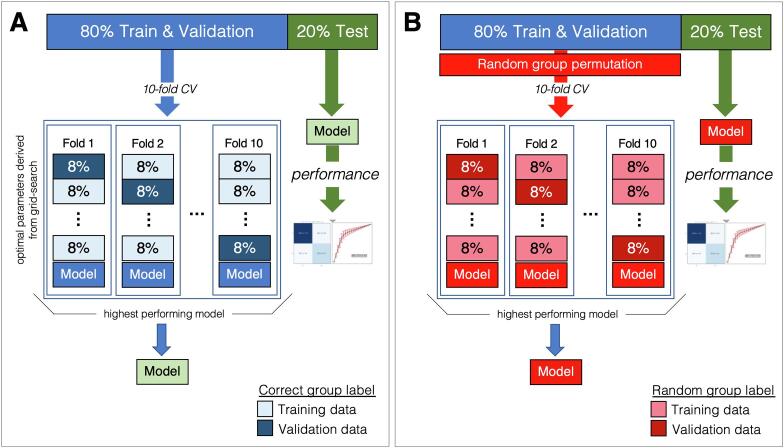
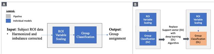
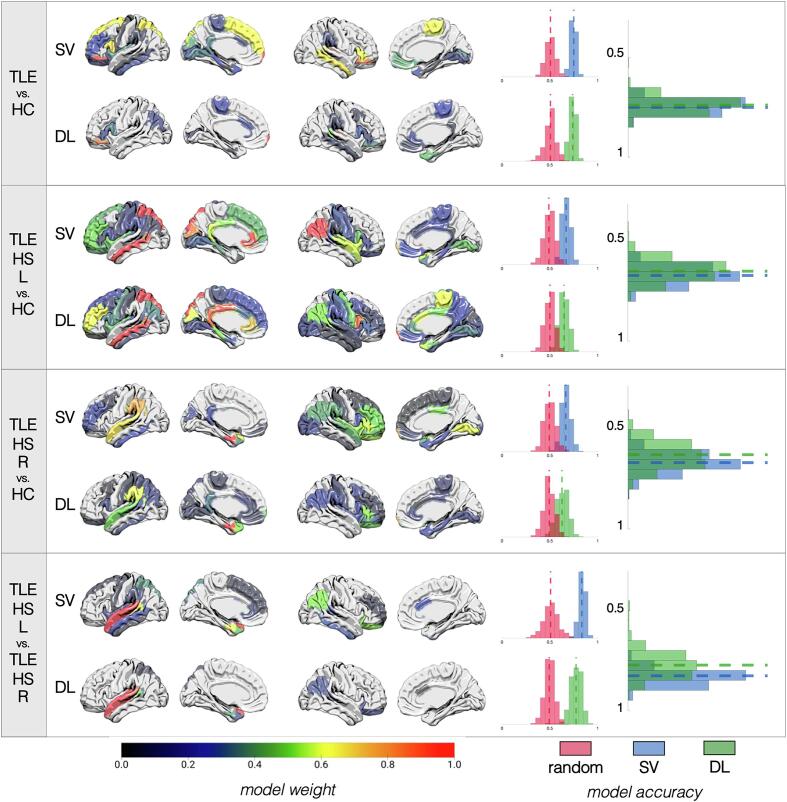
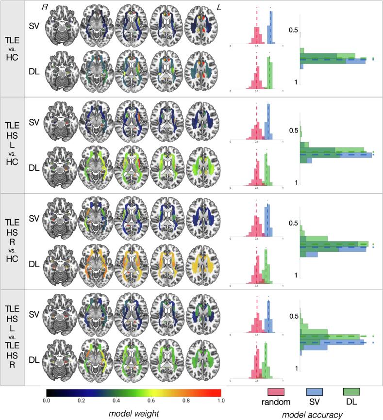
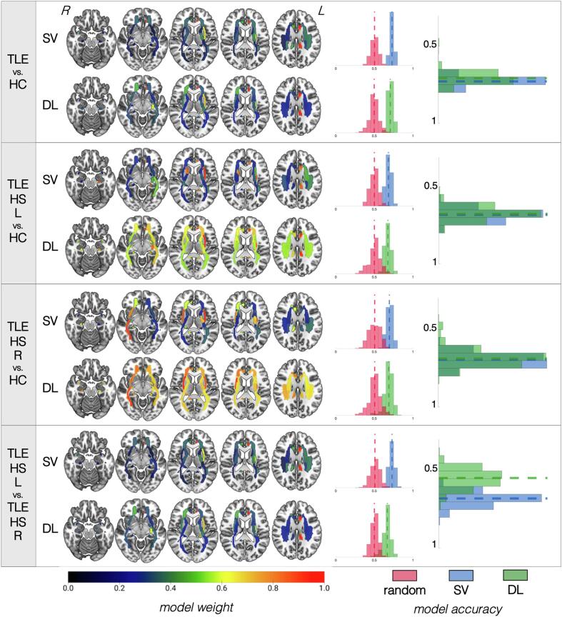
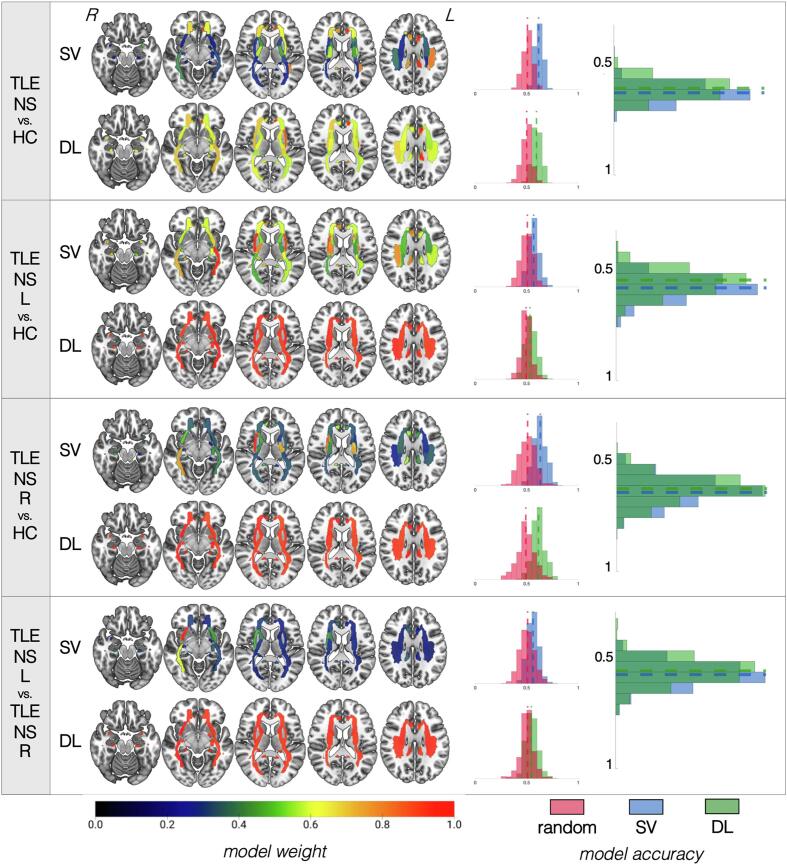
Artificial intelligence has recently gained popularity across different medical fields to aid in the detection of diseases based on pathology samples or medical imaging findings. Brain magnetic resonance imaging (MRI) is a key assessment tool for patients with temporal lobe epilepsy (TLE). The role of machine learning and artificial intelligence to increase detection of brain abnormalities in TLE remains inconclusive. We used support vector machine (SV) and deep learning (DL) models based on region of interest (ROI-based) structural (n = 336) and diffusion (n = 863) brain MRI data from patients with TLE with ("lesional") and without ("non-lesional") radiographic features suggestive of underlying hippocampal sclerosis from the multinational (multi-center) ENIGMA-Epilepsy consortium. Our data showed that models to identify TLE performed better or similar (68-75%) compared to models to lateralize the side of TLE (56-73%, except structural-based) based on diffusion data with the opposite pattern seen for structural data (67-75% to diagnose vs. 83% to lateralize). In other aspects, structural and diffusion-based models showed similar classification accuracies. Our classification models for patients with hippocampal sclerosis were more accurate (68-76%) than models that stratified non-lesional patients (53-62%). Overall, SV and DL models performed similarly with several instances in which SV mildly outperformed DL. We discuss the relative performance of these models with ROI-level data and the implications for future applications of machine learning and artificial intelligence in epilepsy care.
人工智能最近在不同的医学领域得到了广泛应用,可辅助基于病理学样本或医学成像结果对疾病进行检测。脑磁共振成像(MRI)是评估颞叶癫痫(TLE)患者的重要手段。机器学习和人工智能在提高 TLE 脑异常检测中的作用仍存在争议。我们使用支持向量机(SV)和深度学习(DL)模型,基于国际多中心 ENIGMA-Epilepsy 联盟 TLE 患者的感兴趣区(ROI)结构(n=336)和弥散(n=863)脑 MRI 数据,这些患者具有放射学特征提示潜在海马硬化(“病变”)和无(“非病变”)。我们的数据表明,与用于侧化 TLE 侧的模型(56-73%,弥散数据除外,为 67-75%)相比,用于识别 TLE 的模型的性能更好或相似(68-75%),而用于识别 TLE 的模型则相反(67-75%诊断,75%侧化)。在其他方面,结构和弥散模型的分类准确率相似。我们对海马硬化患者的分类模型的准确性(68-76%)高于对非病变患者的分层模型(53-62%)。总体而言,SV 和 DL 模型的性能相似,在某些情况下,SV 略微优于 DL。我们讨论了这些模型与 ROI 级数据的相对性能,以及机器学习和人工智能在癫痫护理中的未来应用的意义。