Wakabayashi Taku, Kamuro Rei, Hashida Noriyasu, Shiraki Nobuhiko, Sakaguchi Hirokazu, Ohguro Nobuyuki, Nishida Kohji
Department of Ophthalmology, Osaka University Graduate School of Medicine, Suita, Japan.
Department of Ophthalmology, Japan Community Health Care Organization Osaka Hospital, Osaka, Japan.
Am J Ophthalmol Case Rep. 2021 Aug 10;23:101188. doi: 10.1016/j.ajoc.2021.101188. eCollection 2021 Sep.
To report a case of acute endophthalmitis and hyphema mimicking pink hypopyon associated with ocular toxocariasis.
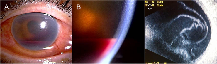
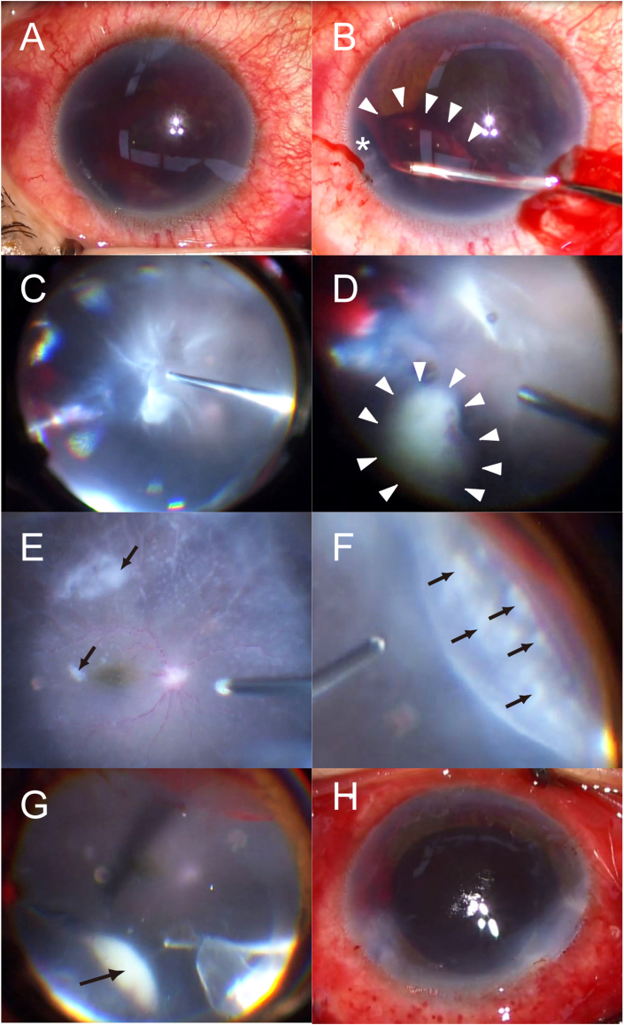
An immunocompetent 56-year-old woman presented to our hospital with a sudden onset and a three-day history of decreased visual acuity in her left eye. There were no known inciting factors for her symptoms; however, she had a history of eating undercooked beef five days prior. On examination, the best-corrected visual acuity of her left eye was light perception and the intraocular pressure was 24 mmHg. Hyphema mimicking pink hypopyon and vitreous opacity suggestive of acute endophthalmitis were observed in her left eye. The patient underwent an emergency pars plana vitrectomy. The intraoperative findings included iridodialysis, severe vitritis, multiple whitish spots on the retina, white sheathed retinal vessels, and whitish peripheral granuloma. The aqueous humor tap and vitreous tap cultures were negative. Blood tests showed elevated eosinophil and total immunoglobulin (Ig) E levels. Enzyme-linked immunosorbent assay of her intraocular fluid showed positive anti-Toxocara canis IgG reactions; the patient was therefore diagnosed with ocular toxocariasis. Subsequent treatment with oral albendazole and prednisone resulted in significant improvement and recovery of visual acuity to 20/12.5.
Acute endophthalmitis with hyphema mimicking pink hypopyon is a rare clinical presentation of ocular toxocariasis. The findings from this case highlight the importance of suspecting ocular toxocariasis if a patient presents with acute endophthalmitis and hyphema accompanied with peripheral granuloma. Early vitrectomy and subsequent treatment with oral albendazole and prednisone can be effective in visual recovery.
报告一例与眼弓蛔虫病相关的急性眼内炎和前房积血,其表现类似粉色积脓性前房积血。
一名免疫功能正常的56岁女性因左眼突然发病且视力下降三天前来我院就诊。其症状无已知诱发因素;然而,她在五天前有食用未煮熟牛肉的病史。检查时,其左眼最佳矫正视力为光感,眼压为24 mmHg。在其左眼观察到类似粉色积脓性前房积血的前房积血以及提示急性眼内炎的玻璃体混浊。患者接受了急诊玻璃体切除术。术中发现包括虹膜根部断离、严重玻璃体炎、视网膜上多个白色斑点、白色鞘膜视网膜血管以及白色周边肉芽肿。房水穿刺和玻璃体穿刺培养均为阴性。血液检查显示嗜酸性粒细胞和总免疫球蛋白(Ig)E水平升高。其眼内液的酶联免疫吸附测定显示抗犬弓蛔虫IgG反应呈阳性;因此,该患者被诊断为眼弓蛔虫病。随后口服阿苯达唑和泼尼松治疗后,视力显著改善,恢复至20/12.5。
伴有类似粉色积脓性前房积血的急性眼内炎是眼弓蛔虫病的一种罕见临床表现。该病例的发现突出了如果患者出现急性眼内炎和前房积血并伴有周边肉芽肿时怀疑眼弓蛔虫病的重要性。早期玻璃体切除术以及随后口服阿苯达唑和泼尼松治疗可有效促进视力恢复。