Prieto-Peña Diana, Castañeda Santos, Martínez-Rodríguez Isabel, Atienza-Mateo Belén, Blanco Ricardo, González-Gay Miguel A
Research Group on Genetic Epidemiology and Atherosclerosis in Systemic Diseases and in Metabolic Bone Diseases of the Musculoskeletal System, IDIVAL, Department of Rheumatology, Hospital Universitario Marqués de Valdecilla, 39008 Santander, Spain.
Department of Rheumatology, Hospital Universitario de La Princesa, IIS-Princesa, 28006 Madrid, Spain.
J Clin Med. 2021 Aug 20;10(16):3704. doi: 10.3390/jcm10163704.
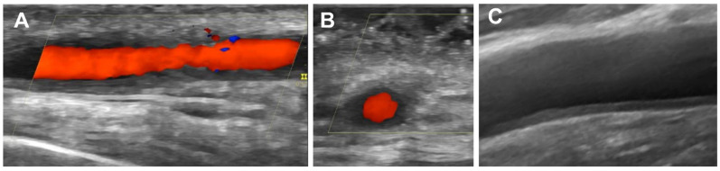
Early recognition of giant cell arteritis (GCA) is crucial to avoid the development of ischemic vascular complications, such as blindness. The classic approach to making the diagnosis of GCA is based on a positive temporal artery biopsy, which is among the criteria proposed by the American College of Rheumatology (ACR) in 1990 to classify a patient as having GCA. However, imaging techniques, particularly ultrasound (US) of the temporal arteries, are increasingly being considered as an alternative for the diagnosis of GCA. Recent recommendations from the European League Against Rheumatism (EULAR) for the use of imaging techniques for large vessel vasculitis (LVV) included US as the first imaging option for the diagnosis of GCA. Furthermore, although the ACR classification criteria are useful in identifying patients with the classic cranial pattern of GCA, they are often inadequate in identifying GCA patients who have the extracranial phenotype of LVV. In this sense, the advent of other imaging techniques, such as magnetic resonance imaging (MRI), computed tomography (CT), and positron emission tomography (PET)/CT, has made it possible to detect the presence of extracranial involvement of the LVV in patients with GCA presenting as refractory rheumatic polymyalgia without cranial ischemic manifestations. Imaging techniques have been the key elements in redefining the diagnostic work-up of GCA. US is currently considered the main imaging modality to improve the early diagnosis of GCA.
早期识别巨细胞动脉炎(GCA)对于避免缺血性血管并发症(如失明)的发生至关重要。诊断GCA的经典方法是基于颞动脉活检阳性,这是美国风湿病学会(ACR)1990年提出的将患者分类为患有GCA的标准之一。然而,成像技术,特别是颞动脉超声(US),越来越被视为诊断GCA的一种替代方法。欧洲抗风湿病联盟(EULAR)最近关于使用成像技术诊断大血管血管炎(LVV)的建议包括将US作为诊断GCA的首选成像方法。此外,尽管ACR分类标准有助于识别具有典型GCA颅部表现的患者,但它们往往不足以识别具有LVV颅外表型的GCA患者。从这个意义上说,其他成像技术,如磁共振成像(MRI)、计算机断层扫描(CT)和正电子发射断层扫描(PET)/CT的出现,使得在表现为难治性风湿性多肌痛且无颅缺血表现的GCA患者中检测LVV颅外受累的情况成为可能。成像技术一直是重新定义GCA诊断检查的关键因素。目前,US被认为是改善GCA早期诊断的主要成像方式。