Pulcini Stefano, Merolle Lucia, Marraccini Chiara, Quartieri Eleonora, Mori Daniele, Schiroli Davide, Berni Pamela, Iotti Barbara, Di Bartolomeo Erminia, Baricchi Roberto, Sala Roberto, Pertinhez Thelma A
Transfusion Medicine Unit, Azienda USL-IRCCS di Reggio Emilia, 42123 Reggio Emilia, Italy.
Department of Medicine and Surgery, University of Parma, 43125 Parma, Italy.
Int J Mol Sci. 2021 Aug 16;22(16):8764. doi: 10.3390/ijms22168764.
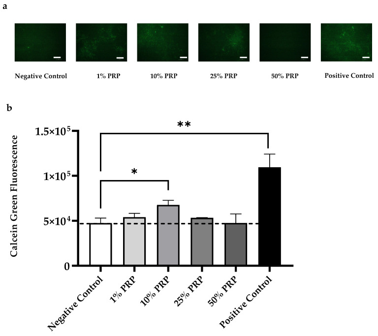
: Platelet-Rich Plasma (PRP) induces bone regeneration; however, there is low evidence supporting its efficacy in bone healing. The lack of a standardized protocol of administration represents the main obstacle to its use in the clinical routine for bone defects' treatment. The purpose of this study was to characterize PRP and elucidate its osteogenic potential. : Platelet count, fibrinogen levels, and growth factors concentration were measured in PRP obtained by four apheresis procedures. HOB-01-C1, a pre-osteocytic cell line, was used to examine the effects of different PRP dilutions (from 1% to 50%) on cell viability, growth, and differentiation. Gene expression of RUNX2, PHEX, COL1A1, and OCN was also assayed. : PRP showed a mean 4.6-fold increase of platelets amount compared to whole blood. Among the 36 proteins evaluated, we found the highest concentrations for PDGF isoforms, EGF, TGF-β and VEGF-D. PDGF-AA positively correlated with platelet counts. In three of the four tested units, 25% PRP induced a growth rate comparable to the positive control (10% FBS); whereas, for all the tested units, 10% PRP treatment sustained differentiation. : This study showed that PRP from apheresis stimulates proliferation and differentiation of pre-osteocyte cells through the release of growth factors from platelets.
富血小板血浆(PRP)可诱导骨再生;然而,支持其在骨愈合中疗效的证据不足。缺乏标准化的给药方案是其在临床常规治疗骨缺损中应用的主要障碍。本研究的目的是对PRP进行表征并阐明其成骨潜力。通过四种单采程序获得的PRP中测量血小板计数、纤维蛋白原水平和生长因子浓度。使用HOB-01-C1,一种前成骨细胞系,来检测不同PRP稀释度(从1%到50%)对细胞活力、生长和分化的影响。还检测了RUNX2、PHEX、COL1A1和OCN的基因表达。与全血相比,PRP显示血小板数量平均增加4.6倍。在评估的36种蛋白质中,我们发现血小板衍生生长因子(PDGF)亚型、表皮生长因子(EGF)、转化生长因子-β(TGF-β)和血管内皮生长因子-D(VEGF-D)的浓度最高。PDGF-AA与血小板计数呈正相关。在四个测试单位中的三个,25%的PRP诱导的生长速率与阳性对照(10%胎牛血清)相当;而对于所有测试单位,10%的PRP处理维持分化。本研究表明,单采获得的PRP通过血小板释放生长因子刺激前成骨细胞的增殖和分化。