Department of Gastroenterology and Hepatology, Mie University Hospital, 2-174 Edobashi, Tsu, Mie, 514-8507, Japan.
Department of Endoscopy, Mie University Hospital, Tsu, Japan.
BMC Gastroenterol. 2021 Aug 28;21(1):336. doi: 10.1186/s12876-021-01898-z.
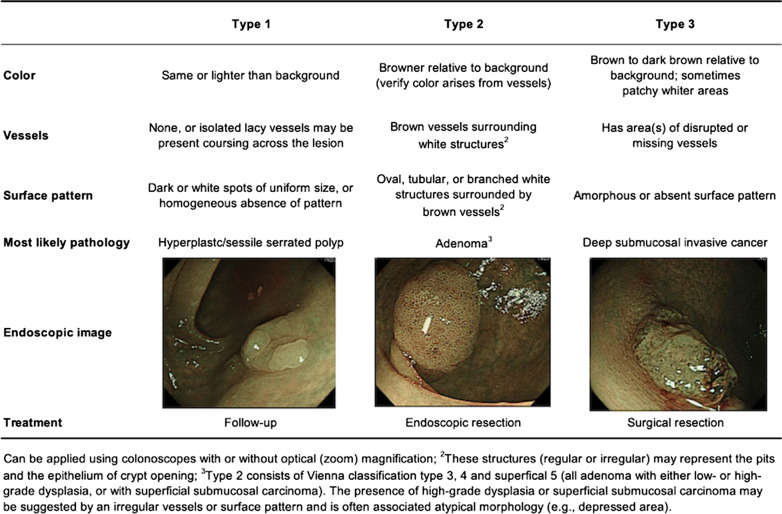
Narrow-band imaging (NBI) highlights the surface structures and vessels of colorectal polyps and is useful for determining the polyp histology. The narrow-band imaging international colorectal endoscopic (NICE) classification is a diagnostic tool for determining colorectal polyp histology based on NBI without optical magnification. In this study, we aimed to investigate the value of each type of the NICE classification for determining colorectal polyp histology using endoscopy data accumulated in a clinical setting.
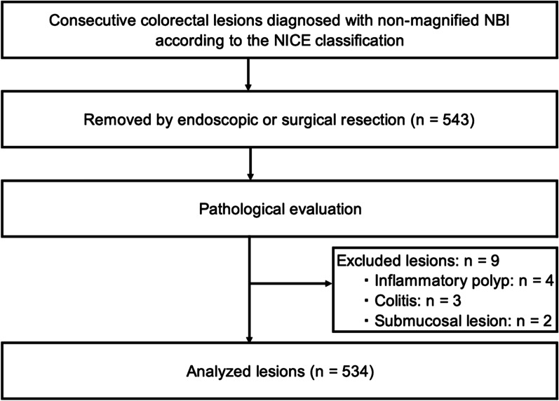
Endoscopy data for 534 colorectal polyps (316 patients) treated at our facility were retrospectively analyzed. First, we investigated the diagnostic performance of each type of the NICE classification for the optical diagnosis of colorectal polyp histology. The procedures were performed by experienced endoscopists using high-definition colonoscopy without optical magnification. Second, inter-observer and intra-observer agreements were assessed after providing experts and non-experts with a short lecture on the NICE classification. Using 50 fine NBI images of colorectal polyps without optical magnification, the inter-observer and intra-observer agreements between five experts and five non-experts were assessed.
The sensitivity, specificity, and accuracy values were 86.0%, 99.6%, and 98.5% for NICE type 1 lesions; 99.2%, 85.2%, and 97.8% for NICE type 2 lesions; and 81.8%, 99.6%, and 99.3% for NICE type 3 lesions, respectively. The inter-observer and intra-observer agreements ranged from substantial to excellent for both experts and non-experts.
The NICE classification had good diagnostic ability in terms of determining the polyp histology and demonstrated a high level of reproducibility among experts and non-experts. Thus, the NICE classification is a useful clinical tool that can be used without optical magnification.
窄带成像(NBI)突出显示结直肠息肉的表面结构和血管,有助于确定息肉的组织学。窄带成像国际结直肠内镜(NICE)分类是一种基于无光学放大的 NBI 确定结直肠息肉组织学的诊断工具。在这项研究中,我们旨在研究在临床环境中积累的内镜数据中,NICE 分类的每种类型用于确定结直肠息肉组织学的价值。
回顾性分析了在我院治疗的 534 个结直肠息肉(316 例患者)的内镜数据。首先,我们研究了 NICE 分类的每种类型对结直肠息肉组织学的光学诊断的诊断性能。这些程序是由经验丰富的内镜医师在没有光学放大的高清结肠镜下进行的。其次,在为专家和非专家提供关于 NICE 分类的简短讲座后,评估了观察者间和观察者内的一致性。使用 50 张无光学放大的结直肠息肉的精细 NBI 图像,评估了五名专家和五名非专家之间的观察者间和观察者内一致性。
对于 NICE 1 型病变,灵敏度、特异性和准确性值分别为 86.0%、99.6%和 98.5%;对于 NICE 2 型病变,分别为 99.2%、85.2%和 97.8%;对于 NICE 3 型病变,分别为 81.8%、99.6%和 99.3%。专家和非专家的观察者间和观察者内一致性均从中等到极好。
NICE 分类在确定息肉组织学方面具有良好的诊断能力,并且在专家和非专家之间具有高度的可重复性。因此,NICE 分类是一种有用的临床工具,可以在没有光学放大的情况下使用。