• 文献检索
  • 文档翻译
  • 深度研究
  • 学术资讯
  • Suppr Zotero 插件Zotero 插件
  • 邀请有礼
  • 套餐&价格
  • 历史记录
应用&插件
Suppr Zotero 插件Zotero 插件浏览器插件Mac 客户端Windows 客户端微信小程序
定价
高级版会员购买积分包购买API积分包
服务
文献检索文档翻译深度研究API 文档MCP 服务
关于我们
关于 Suppr公司介绍联系我们用户协议隐私条款
关注我们

Suppr 超能文献

核心技术专利:CN118964589B侵权必究
粤ICP备2023148730 号-1Suppr @ 2026

文献检索

告别复杂PubMed语法,用中文像聊天一样搜索,搜遍4000万医学文献。AI智能推荐,让科研检索更轻松。

立即免费搜索

文件翻译

保留排版,准确专业,支持PDF/Word/PPT等文件格式,支持 12+语言互译。

免费翻译文档

深度研究

AI帮你快速写综述,25分钟生成高质量综述,智能提取关键信息,辅助科研写作。

立即免费体验

正常和坏血病猴的视网膜光损伤

Retinal photic injury in normal and scorbutic monkeys.

作者信息

Tso M O

机构信息

University of Illinois College of Medicine at Chicago.

出版信息

Trans Am Ophthalmol Soc. 1987;85:498-556.

PMID:3447341
原文链接:https://pmc.ncbi.nlm.nih.gov/articles/PMC1298790/
Abstract

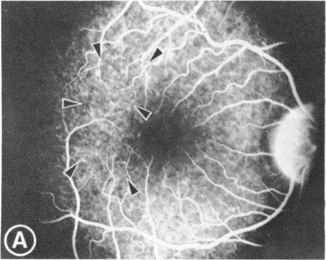
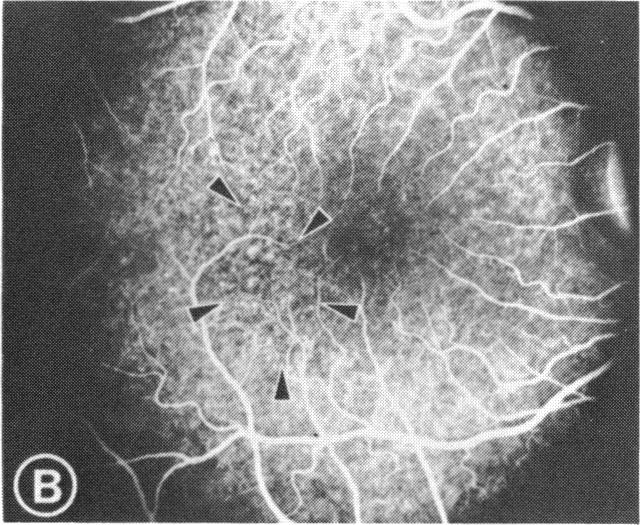
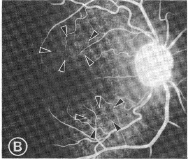
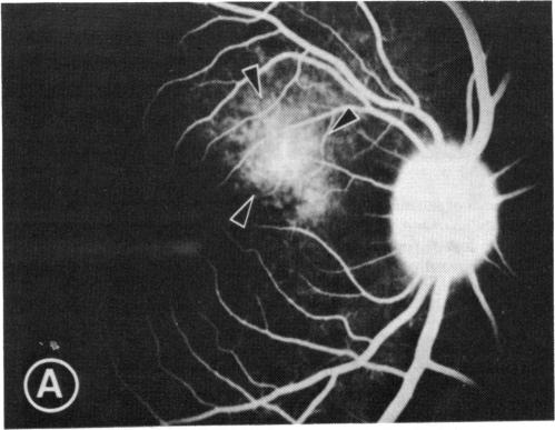
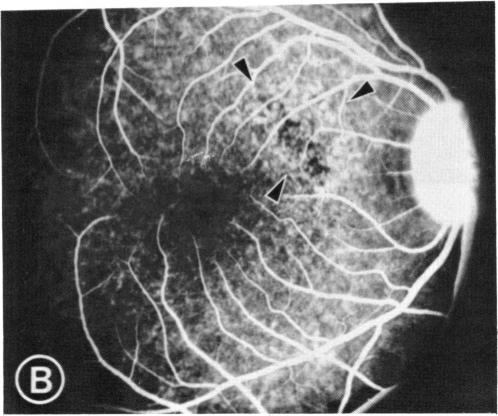
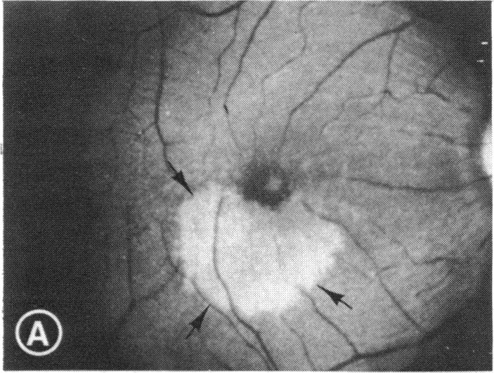
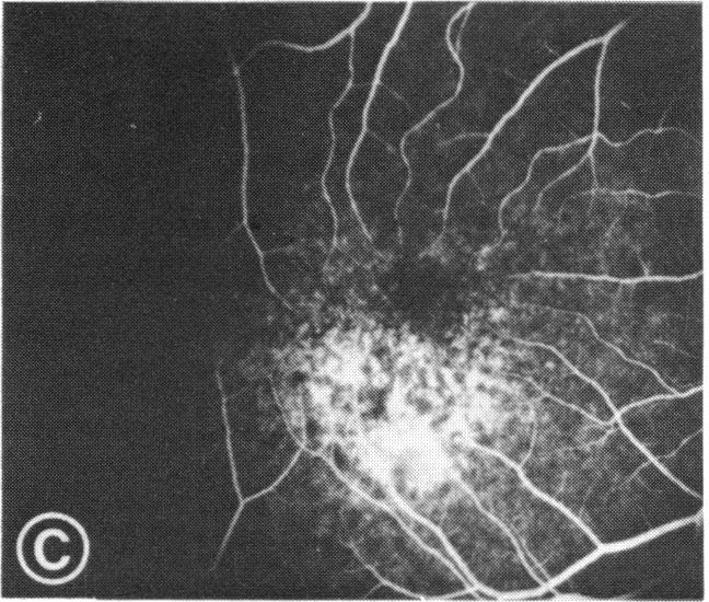
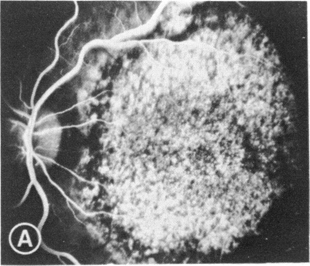
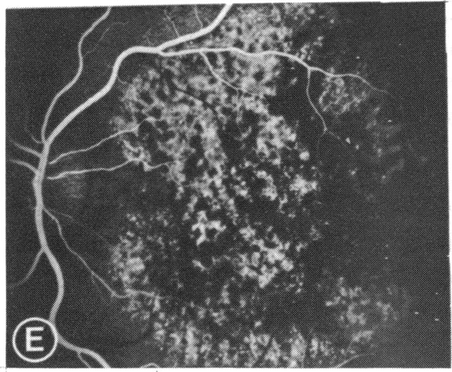
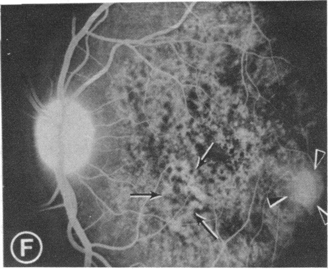
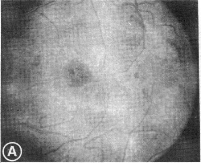
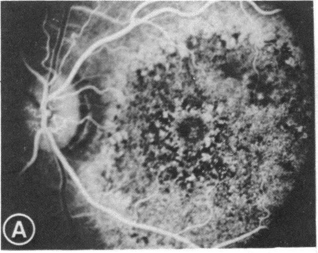
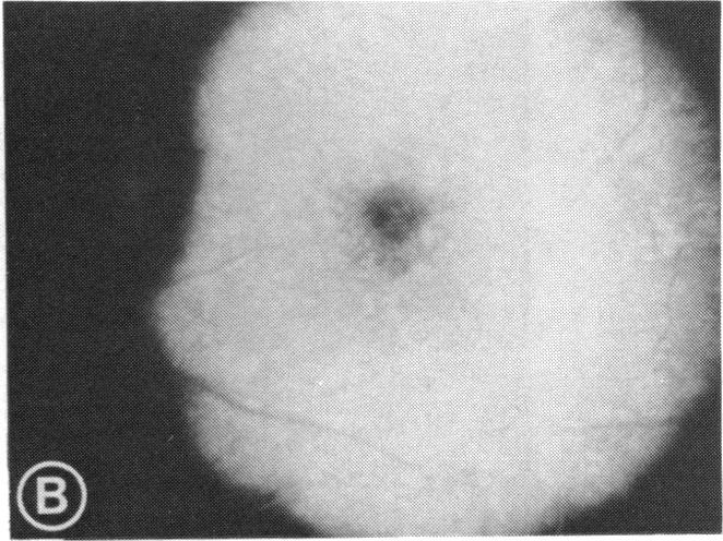
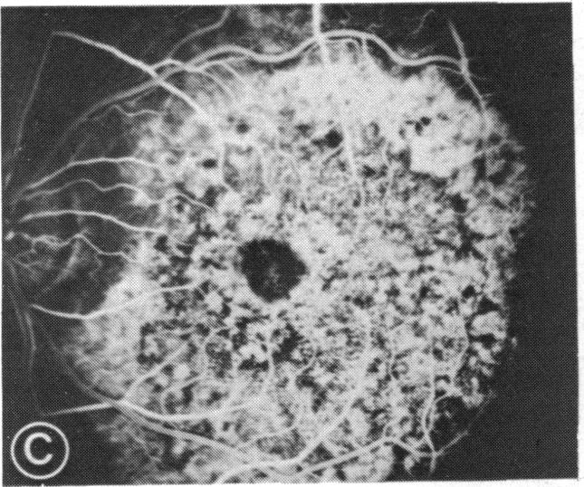
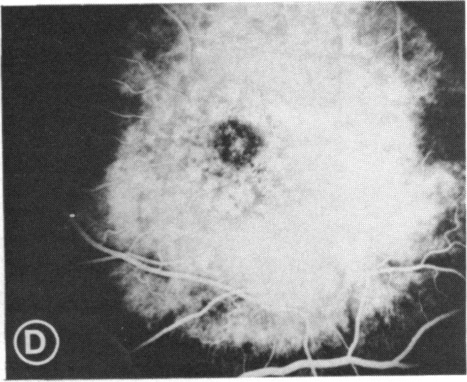
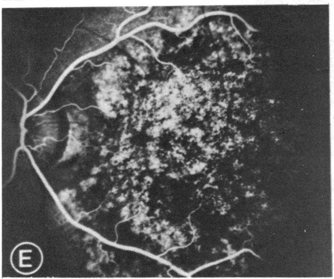
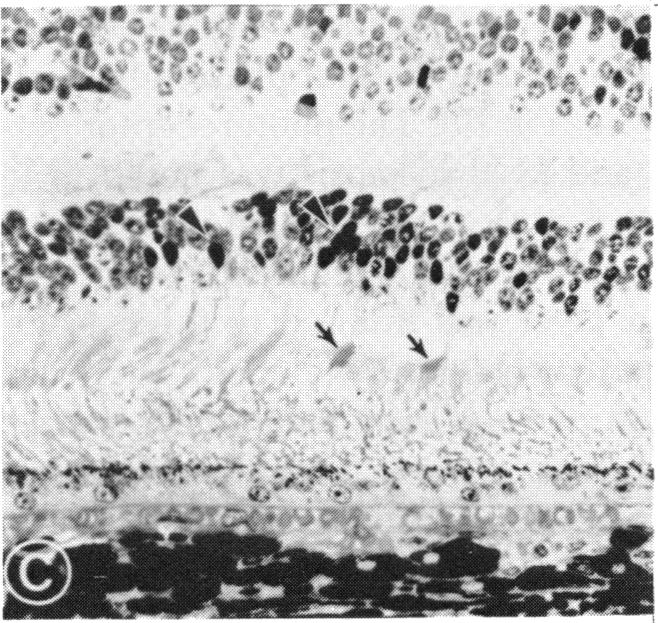
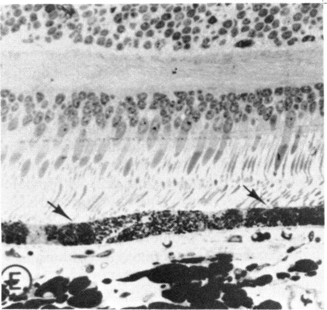
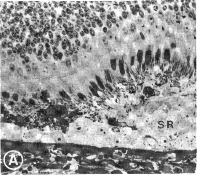
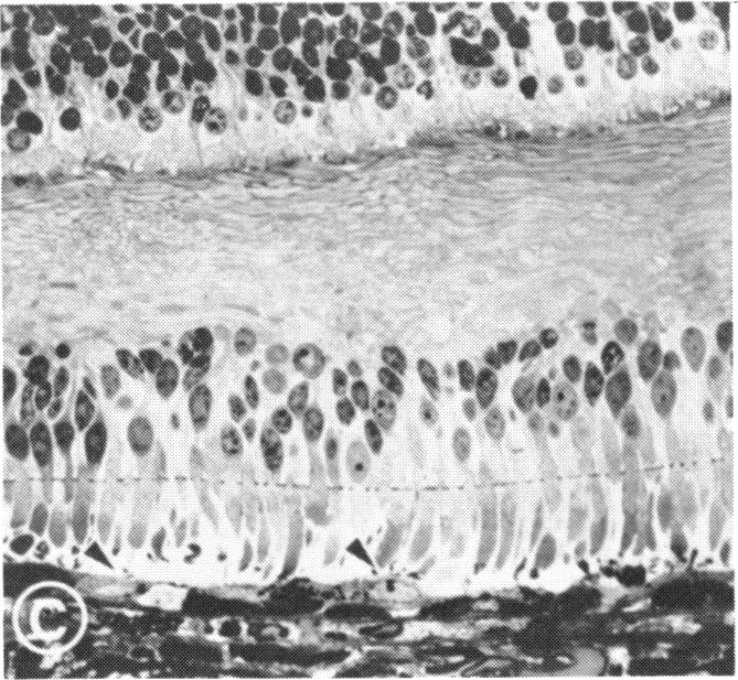
Mild and severe retinal photic injuries were inflicted on 22 eyes of seven monkeys fed a vitamin C-deficient diet and four monkeys given a vitamin C-enriched diet. The retinal lesions were studied by fundus examination, fluorescein angiography, and light and electron microscopy. While the general cellular response to photic injury in the retina of scorbutic animals was not different qualitatively from that in the normal animals, scurvy appeared to cause more severe tissue damage, an exaggerated repair response, and more advanced retinal degeneration. In the four groups of eyes, representing mild and severe photic injury in normal and scorbutic animals, a continuous spectrum of changes was produced. The least damage occurred from mild photic injury in the normal animals, and the most detrimental insult resulted from severe photic injury in the scorbutic animals. We propose that the basic mechanism by which ascorbate mitigates retinal photic injury depends on its redox properties. Ascorbate functions as an antioxidant in the retina. It scavenges superoxide radicals and hydroxyl radicals, quenches singlet oxygen, and reduces hydrogen peroxide, all of which are formed in retinal photic injury. This hypothesis provides an explanation for the high level of ascorbate in the retina. The pathogenetic mechanisms that correspond to the three distinct phases of pathologic changes observed in retinal photic injury are characterized. In phase 1, single oxygen is generated in a photodynamic reaction that damages the photoreceptor elements and pigment epithelium. In phase 2, macrophages attracted from the systemic circulation invade the subretinal space, and a photo-oxidative reaction generates superoxide radicals, hydrogen peroxide, and hydroxyl radicals. These free radicals attack the photoreceptor cells and pigment epithelium to cause further retinal damage. In phase 3, macrophages remain in the subretinal space for as long as 8 months after injury, causing persistent disruption of the blood-retinal barrier. The photo-oxidative reaction appears to linger, resulting in chronic retinal degeneration. It is hypothesized that in some forms of age-related macular degeneration, patients suffer from repeated mild photic insult throughout their lifetime. Aging has been associated with subclinical scurvy, which leads to even greater susceptibility to photic injury. Although ascorbate moderates many biochemical functions of the body and helps the retina ameliorate photo-oxidative injury, it should be regarded as a nutritional supplement to maintain health when consumed in appropriate amounts and not as a therapeutic agent for the treatment of severe insults.

摘要

对7只喂食缺乏维生素C饮食的猴子的22只眼睛以及4只喂食富含维生素C饮食的猴子的眼睛造成轻度和重度视网膜光损伤。通过眼底检查、荧光素血管造影以及光镜和电镜对视网膜病变进行研究。虽然坏血病动物视网膜对光损伤的一般细胞反应在性质上与正常动物没有差异,但坏血病似乎会导致更严重的组织损伤、过度的修复反应以及更严重的视网膜变性。在四组眼睛中,分别代表正常和坏血病动物的轻度和重度光损伤,产生了一系列连续的变化。损伤最小的是正常动物的轻度光损伤,而最具破坏性的损伤是坏血病动物的重度光损伤。我们提出抗坏血酸减轻视网膜光损伤的基本机制取决于其氧化还原特性。抗坏血酸在视网膜中起抗氧化剂的作用。它清除超氧自由基和羟自由基,淬灭单线态氧,并还原过氧化氢,所有这些都是在视网膜光损伤中形成的。这一假设为视网膜中高水平的抗坏血酸提供了解释。描述了与视网膜光损伤中观察到的三个不同病理变化阶段相对应的发病机制。在第1阶段,光动力反应中产生单线态氧,损害光感受器元件和色素上皮。在第2阶段,从全身循环吸引来的巨噬细胞侵入视网膜下间隙,光氧化反应产生超氧自由基、过氧化氢和羟自由基。这些自由基攻击光感受器细胞和色素上皮,导致进一步的视网膜损伤。在第3阶段,巨噬细胞在损伤后长达8个月的时间内一直留在视网膜下间隙,导致血视网膜屏障持续破坏。光氧化反应似乎持续存在,导致慢性视网膜变性。据推测,在某些形式的年龄相关性黄斑变性中,患者一生中会遭受反复的轻度光损伤。衰老与亚临床坏血病有关,这导致对光损伤的易感性更高。虽然抗坏血酸调节身体的许多生化功能并帮助视网膜减轻光氧化损伤,但当适量摄入时,它应被视为维持健康的营养补充剂,而不是用于治疗严重损伤的治疗剂。

https://cdn.ncbi.nlm.nih.gov/pmc/blobs/5d79/1298790/b0bd0cb53081/taos00014-0544-e.jpg
https://cdn.ncbi.nlm.nih.gov/pmc/blobs/5d79/1298790/86628ae72373/taos00014-0533-a.jpg
https://cdn.ncbi.nlm.nih.gov/pmc/blobs/5d79/1298790/5c7b20880ac0/taos00014-0533-b.jpg
https://cdn.ncbi.nlm.nih.gov/pmc/blobs/5d79/1298790/5c15b64bc5fa/taos00014-0533-c.jpg
https://cdn.ncbi.nlm.nih.gov/pmc/blobs/5d79/1298790/03deb779e3db/taos00014-0533-d.jpg
https://cdn.ncbi.nlm.nih.gov/pmc/blobs/5d79/1298790/585d34b29a73/taos00014-0534-a.jpg
https://cdn.ncbi.nlm.nih.gov/pmc/blobs/5d79/1298790/2c6a53e27e43/taos00014-0534-b.jpg
https://cdn.ncbi.nlm.nih.gov/pmc/blobs/5d79/1298790/a3c86c9f8570/taos00014-0536-a.jpg
https://cdn.ncbi.nlm.nih.gov/pmc/blobs/5d79/1298790/a59d6cc42e4c/taos00014-0536-b.jpg
https://cdn.ncbi.nlm.nih.gov/pmc/blobs/5d79/1298790/1bdf7b0ee123/taos00014-0536-c.jpg
https://cdn.ncbi.nlm.nih.gov/pmc/blobs/5d79/1298790/e3d6fb3ff791/taos00014-0536-d.jpg
https://cdn.ncbi.nlm.nih.gov/pmc/blobs/5d79/1298790/e2174fbc4eb7/taos00014-0536-e.jpg
https://cdn.ncbi.nlm.nih.gov/pmc/blobs/5d79/1298790/298bd9a025ff/taos00014-0537-a.jpg
https://cdn.ncbi.nlm.nih.gov/pmc/blobs/5d79/1298790/369fc83b66ec/taos00014-0537-b.jpg
https://cdn.ncbi.nlm.nih.gov/pmc/blobs/5d79/1298790/c84c1714e132/taos00014-0537-c.jpg
https://cdn.ncbi.nlm.nih.gov/pmc/blobs/5d79/1298790/6dfe6eca91b0/taos00014-0537-d.jpg
https://cdn.ncbi.nlm.nih.gov/pmc/blobs/5d79/1298790/050bbea9a4bf/taos00014-0537-e.jpg
https://cdn.ncbi.nlm.nih.gov/pmc/blobs/5d79/1298790/6188d059c345/taos00014-0537-f.jpg
https://cdn.ncbi.nlm.nih.gov/pmc/blobs/5d79/1298790/b37ed528c8e8/taos00014-0538-a.jpg
https://cdn.ncbi.nlm.nih.gov/pmc/blobs/5d79/1298790/da03da9b4afb/taos00014-0538-b.jpg
https://cdn.ncbi.nlm.nih.gov/pmc/blobs/5d79/1298790/ab77435698ce/taos00014-0539-a.jpg
https://cdn.ncbi.nlm.nih.gov/pmc/blobs/5d79/1298790/e8c3a314eb89/taos00014-0539-b.jpg
https://cdn.ncbi.nlm.nih.gov/pmc/blobs/5d79/1298790/af16beb8dfeb/taos00014-0539-c.jpg
https://cdn.ncbi.nlm.nih.gov/pmc/blobs/5d79/1298790/7a6bd4f16584/taos00014-0539-d.jpg
https://cdn.ncbi.nlm.nih.gov/pmc/blobs/5d79/1298790/5f0ae84cf712/taos00014-0539-e.jpg
https://cdn.ncbi.nlm.nih.gov/pmc/blobs/5d79/1298790/7bec79bfb427/taos00014-0539-f.jpg
https://cdn.ncbi.nlm.nih.gov/pmc/blobs/5d79/1298790/97ae9efc59e4/taos00014-0540-a.jpg
https://cdn.ncbi.nlm.nih.gov/pmc/blobs/5d79/1298790/01b5e6670da7/taos00014-0540-b.jpg
https://cdn.ncbi.nlm.nih.gov/pmc/blobs/5d79/1298790/3f99d5a397c1/taos00014-0540-c.jpg
https://cdn.ncbi.nlm.nih.gov/pmc/blobs/5d79/1298790/4a66b95dc4fa/taos00014-0542-a.jpg
https://cdn.ncbi.nlm.nih.gov/pmc/blobs/5d79/1298790/284674948825/taos00014-0542-c.jpg
https://cdn.ncbi.nlm.nih.gov/pmc/blobs/5d79/1298790/949e735e193e/taos00014-0542-d.jpg
https://cdn.ncbi.nlm.nih.gov/pmc/blobs/5d79/1298790/2e73c78fc8ee/taos00014-0542-e.jpg
https://cdn.ncbi.nlm.nih.gov/pmc/blobs/5d79/1298790/d163174c3dad/taos00014-0542-f.jpg
https://cdn.ncbi.nlm.nih.gov/pmc/blobs/5d79/1298790/70542e357711/taos00014-0543-a.jpg
https://cdn.ncbi.nlm.nih.gov/pmc/blobs/5d79/1298790/b58e0460795f/taos00014-0543-c.jpg
https://cdn.ncbi.nlm.nih.gov/pmc/blobs/5d79/1298790/9da71297e672/taos00014-0543-d.jpg
https://cdn.ncbi.nlm.nih.gov/pmc/blobs/5d79/1298790/c61d92381758/taos00014-0544-b.jpg
https://cdn.ncbi.nlm.nih.gov/pmc/blobs/5d79/1298790/b0bd0cb53081/taos00014-0544-e.jpg

相似文献

1
Retinal photic injury in normal and scorbutic monkeys.正常和坏血病猴的视网膜光损伤
Trans Am Ophthalmol Soc. 1987;85:498-556.
2
Effect of photic injury on the retinal tissues.光损伤对视网膜组织的影响。
Ophthalmology. 1983 Aug;90(8):952-63. doi: 10.1016/s0161-6420(83)80023-2.
3
Amelioration of photic injury in rat retina by ascorbic acid: a histopathologic study.
Invest Ophthalmol Vis Sci. 1985 Nov;26(11):1589-98.
4
Increased expression of ceruloplasmin in the retina following photic injury.光损伤后视网膜中铜蓝蛋白表达增加。
Mol Vis. 2003 Apr 30;9:151-8.
5
[Retinal reactions to intense light. I. Threshold lesions. Experimental, morphological and clinical studies of pathological and therapeutic effects of laser and white light].[视网膜对强光的反应。I. 阈值性病变。激光和白光的病理及治疗作用的实验、形态学和临床研究]
Adv Ophthalmol. 1975;31:159-232.
6
[A new approach for studying the retinal and choroidal circulation].[一种研究视网膜和脉络膜循环的新方法]
Nippon Ganka Gakkai Zasshi. 2004 Dec;108(12):836-61; discussion 862.
7
Retinal damage produced by intraocular fiber optic light.眼内光纤光造成的视网膜损伤。
Am J Ophthalmol. 1978 Apr;85(4):519-37. doi: 10.1016/s0002-9394(14)75250-x.
8
The broad-spectrum chemokine inhibitor NR58-3.14.3 modulates macrophage-mediated inflammation in the diseased retina.广谱趋化因子抑制剂NR58-3.14.3可调节病变视网膜中巨噬细胞介导的炎症反应。
J Neuroinflammation. 2016 Feb 24;13:47. doi: 10.1186/s12974-016-0514-x.
9
Effects of repeated photic exposures on the monkey macula.
Arch Ophthalmol. 1990 May;108(5):727-33. doi: 10.1001/archopht.1990.01070070113047.
10
Reduced and oxidized ascorbates in guinea pig retina under normal and light-exposed conditions.正常和光照条件下豚鼠视网膜中的还原型和氧化型抗坏血酸盐
Invest Ophthalmol Vis Sci. 1983 Jul;24(7):862-7.

引用本文的文献

1
Protective effect of alpha-mangostin against oxidative stress induced-retinal cell death.α-山竹黄酮对氧化应激诱导的视网膜细胞死亡的保护作用。
Sci Rep. 2016 Feb 18;6:21018. doi: 10.1038/srep21018.
2
Influence of blue light on photoreceptors in a live retinal explant system.蓝光对活体视网膜外植体系统中光感受器的影响。
Mol Vis. 2011 Apr 8;17:876-84.
3
Retinal light toxicity.视网膜光毒性。

本文引用的文献

1
The Rôle of Ascorbic Acid (Vitamin C) in the Secretion of the Intra-Ocular Fluid.抗坏血酸(维生素C)在眼房水分泌中的作用
Trans Am Ophthalmol Soc. 1939;37:310-35.
2
The free radical property of melanins.黑色素的自由基特性。
Arch Biochem Biophys. 1960 Feb;86:225-30. doi: 10.1016/0003-9861(60)90409-4.
3
Influence of local ascorbic acid concentration on collagenous tissue healing in the cornea.局部抗坏血酸浓度对角膜胶原组织愈合的影响。
Eye (Lond). 2011 Jan;25(1):1-14. doi: 10.1038/eye.2010.149. Epub 2010 Oct 29.
4
Retinal light damage: mechanisms and protection.视网膜光损伤:机制与保护。
Prog Retin Eye Res. 2010 Mar;29(2):113-34. doi: 10.1016/j.preteyeres.2009.11.004. Epub 2009 Dec 3.
5
Pro-inflammatory cytokines increase reactive oxygen species through mitochondria and NADPH oxidase in cultured RPE cells.促炎细胞因子通过线粒体和NADPH氧化酶增加培养的视网膜色素上皮(RPE)细胞中的活性氧。
Exp Eye Res. 2007 Oct;85(4):462-72. doi: 10.1016/j.exer.2007.06.013. Epub 2007 Jun 27.
6
Effects of ascorbic acid on UV light-mediated photoreceptor damage in isolated rat retina.抗坏血酸对离体大鼠视网膜中紫外线介导的光感受器损伤的影响。
Exp Eye Res. 2007 Mar;84(3):537-43. doi: 10.1016/j.exer.2006.11.005. Epub 2007 Jan 12.
7
How much blue light should an IOL transmit?人工晶状体应传输多少蓝光?
Br J Ophthalmol. 2003 Dec;87(12):1523-9. doi: 10.1136/bjo.87.12.1523.
8
Nitric oxide and lipid peroxidation are increased and associated with decreased antioxidant enzyme activities in patients with age-related macular degeneration.在年龄相关性黄斑变性患者中,一氧化氮和脂质过氧化增加,并与抗氧化酶活性降低有关。
Doc Ophthalmol. 2003 Mar;106(2):129-36. doi: 10.1023/a:1022512402811.
9
Retinal pigment epithelial acid lipase activity and lipoprotein receptors: effects of dietary omega-3 fatty acids.视网膜色素上皮酸脂肪酶活性与脂蛋白受体:膳食ω-3脂肪酸的影响
Trans Am Ophthalmol Soc. 2002;100:301-38.
10
Visible light and risk of age-related macular degeneration.可见光与年龄相关性黄斑变性风险
Trans Am Ophthalmol Soc. 1990;88:163-73; discussion 173-8.
Br J Ophthalmol. 1955 Apr;39(4):204-14. doi: 10.1136/bjo.39.4.204.
4
Changes in the vitreous body in scurvy; studies on guinea pigs in vivo. I. The biopolymers of the vitreous body.坏血病时玻璃体的变化;对豚鼠的体内研究。I. 玻璃体的生物聚合物
Acta Ophthalmol (Copenh). 1957;35(4):336-60. doi: 10.1111/j.1755-3768.1957.tb07696.x.
5
Solar chorioretinal burn.日光性脉络膜视网膜灼伤
Am J Ophthalmol. 1956 Jun;41(6):1048-53. doi: 10.1016/0002-9394(56)91056-x.
6
Ascorbic acid as a test substance for measuring relative changes in the rate of plasma flow through the ciliary processes. IV. The effect of carotid ligation and cervical sympathectomy in guinea-pigs on the ascorbic acid content of the aqueous humour at varying plasma levels.抗坏血酸作为一种测试物质,用于测量通过睫状体的血浆流速的相对变化。IV. 豚鼠颈动脉结扎和颈交感神经切除对不同血浆水平时房水中抗坏血酸含量的影响。
Acta Physiol Scand. 1952 Sep 10;26(2-3):130-47. doi: 10.1111/j.1748-1716.1952.tb00896.x.
7
Ascorbic acid as a test substance for measuring relative changes in the rate of plasma flow through the ciliary processes. III. The effect of pre-ganglionic section of the cervical sympathetic in rabbits on the ascorbic acid content of the aqueous humour at varying plasma levels.
Acta Physiol Scand. 1952 Jul 17;26(1):79-85. doi: 10.1111/j.1748-1716.1952.tb00893.x.
8
Aging and degeneration of the human macula. 1. Outer nuclear layer and photoreceptors.人类黄斑的衰老与退变。1. 外核层与光感受器。
Br J Ophthalmol. 1981 Jan;65(1):23-8. doi: 10.1136/bjo.65.1.23.
9
Lipofuscin of human retinal pigment epithelium.人视网膜色素上皮的脂褐素。
Am J Ophthalmol. 1980 Dec;90(6):783-91. doi: 10.1016/s0002-9394(14)75193-1.
10
The Framingham Eye Study monograph: An ophthalmological and epidemiological study of cataract, glaucoma, diabetic retinopathy, macular degeneration, and visual acuity in a general population of 2631 adults, 1973-1975.《弗雷明汉姆眼科研究专论》:对2631名成年人组成的普通人群进行的白内障、青光眼、糖尿病视网膜病变、黄斑变性及视力的眼科与流行病学研究,1973 - 1975年。
Surv Ophthalmol. 1980 May-Jun;24(Suppl):335-610.