Zhe Xia, Chen Li, Zhang Dongsheng, Tang Min, Gao Jie, Ai Kai, Liu Weijun, Lei Xiaoyan, Zhang Xiaoling
Department of MRI, Shaanxi Provincial People's Hospital, Xi'an, China.
Department of Neurology, Shaanxi Provincial People's Hospital, Xi'an, China.
Front Hum Neurosci. 2021 Aug 16;15:717130. doi: 10.3389/fnhum.2021.717130. eCollection 2021.
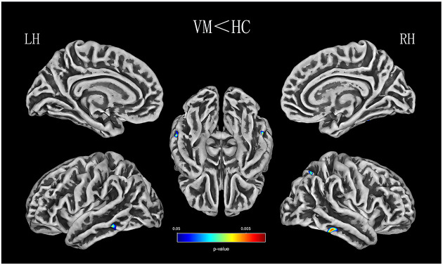
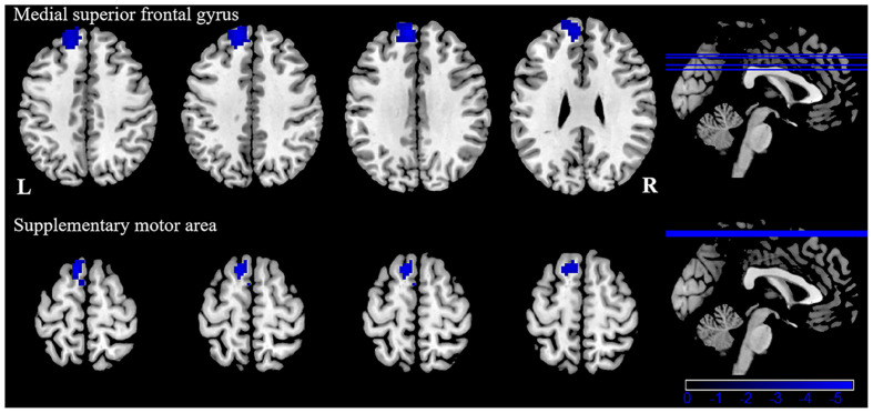
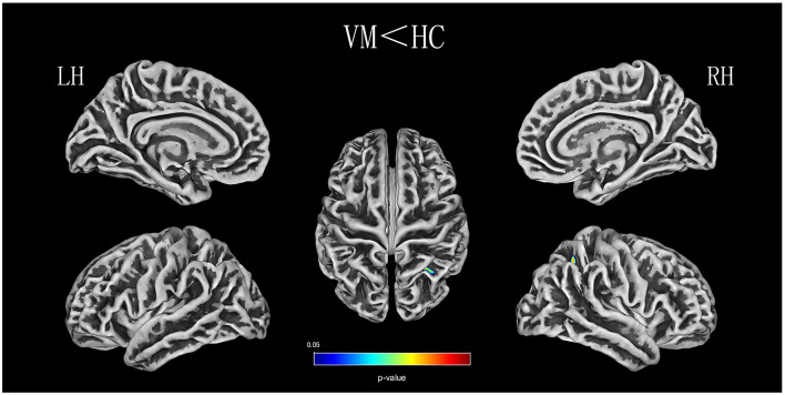
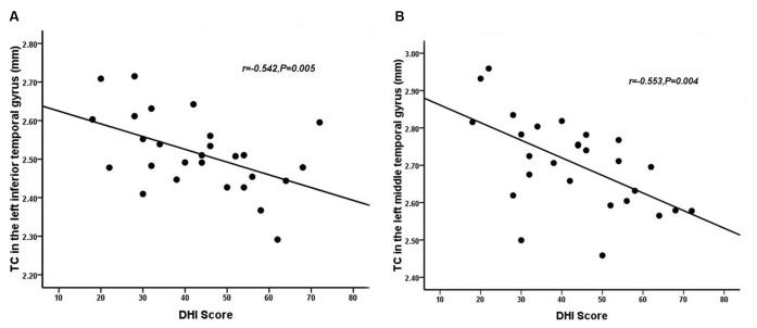
: Increasing evidence suggests that the temporal and parietal lobes are associated with multisensory integration and vestibular migraine. However, temporal and parietal lobe structural and functional connectivity (FC) changes related to vestibular migraine need to be further investigated. : Twenty-five patients with vestibular migraine (VM) and 27 age- and sex- matched healthy controls participated in this study. Participants completed standardized questionnaires assessing migraine and vertigo-related clinical features. Cerebral cortex characteristics [i.e., thickness (CT), fractal dimension (FD), sulcus depth (SD), and the gyrification index (GI)] were evaluated using an automated Computational Anatomy Toolbox (CAT12). Regions with significant differences were used in a seed-based comparison of resting-state FC conducted with DPABI. The relationship between changes in cortical characteristics or FC and clinical features was also analyzed in the patients with VM. : Relative to controls, patients with VM showed significantly thinner CT in the bilateral inferior temporal gyrus, left middle temporal gyrus, and the right superior parietal lobule. A shallower SD was observed in the right superior and inferior parietal lobule. FD and GI did not differ significantly between the two groups. A negative correlation was found between CT in the right inferior temporal gyrus, as well as the left middle temporal gyrus, and the Dizziness Handicap Inventory (DHI) score in VM patients. Furthermore, patients with VM exhibited weaker FC between the left inferior/middle temporal gyrus and the left medial superior frontal gyrus, supplementary motor area. : Our data revealed cortical structural and resting-state FC abnormalities associated with multisensory integration, contributing to a lower quality of life. These observations suggest a role for multisensory integration in patients with VM pathophysiology. Future research should focus on using a task-based fMRI to measure multisensory integration.
越来越多的证据表明,颞叶和顶叶与多感觉整合及前庭性偏头痛有关。然而,与前庭性偏头痛相关的颞叶和顶叶结构及功能连接(FC)变化仍需进一步研究。25例前庭性偏头痛(VM)患者和27例年龄及性别匹配的健康对照者参与了本研究。参与者完成了评估偏头痛和眩晕相关临床特征的标准化问卷。使用自动化的计算解剖工具箱(CAT12)评估大脑皮质特征[即厚度(CT)、分形维数(FD)、脑沟深度(SD)和脑回指数(GI)]。在基于种子点的静息态FC比较中,使用DPABI对有显著差异的区域进行分析。还分析了VM患者皮质特征或FC变化与临床特征之间的关系。相对于对照组,VM患者双侧颞下回、左侧颞中回和右侧顶上小叶的CT显著变薄。右侧顶叶上下小叶的SD较浅。两组之间FD和GI无显著差异。VM患者右侧颞下回以及左侧颞中回的CT与头晕残障量表(DHI)评分呈负相关。此外,VM患者左侧颞下/中回与左侧额上回内侧、辅助运动区之间的FC较弱。我们的数据揭示了与多感觉整合相关的皮质结构和静息态FC异常,这导致了生活质量下降。这些观察结果表明多感觉整合在VM病理生理学中起作用。未来的研究应侧重于使用基于任务的功能磁共振成像来测量多感觉整合。