Gumru Birsay, Akkitap Melda Pelin, Deveci Sevilay, Idman Ender
Department of Oral and Maxillofacial Radiology, Faculty of Dentistry, Marmara University, Istanbul, Turkey.
J Dent Sci. 2021 Oct;16(4):1154-1161. doi: 10.1016/j.jds.2021.03.009. Epub 2021 Apr 7.
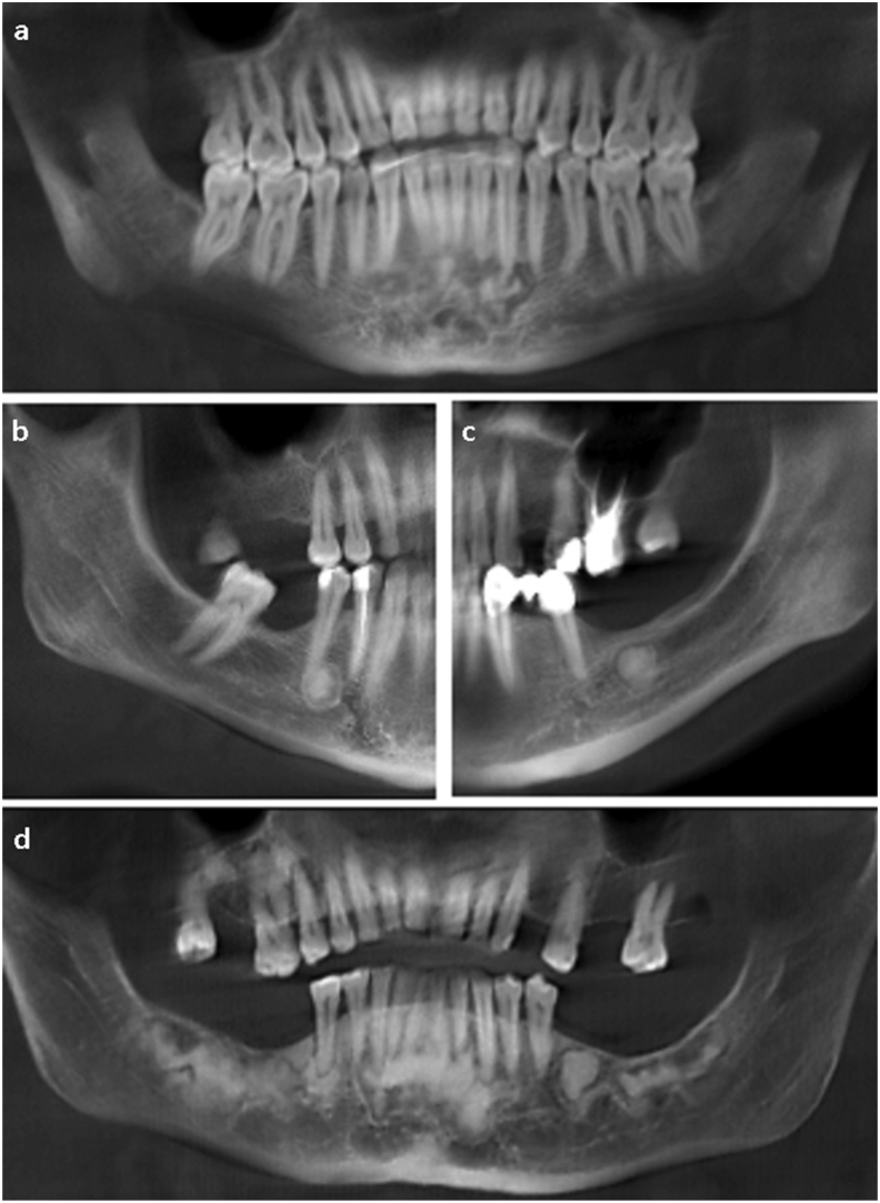
BACKGROUND/PURPOSE: Radiological examination is indispensable in the diagnosis and follow-up of cemento-osseous dysplasia (COD). The aim of this retrospective study was to describe a series of COD cases, identify the frequencies of COD subtypes, and investigate the demographic and radiological characteristics in relation to subtypes.
Cone beam computed tomography (CBCT) images/reports of patients with a diagnosis of COD were included in the study. The data collected included information on the age, sex, subtype of COD, location of COD, and region involved. Information regarding the internal density, effects on surrounding structures, and presence of concomitant lesions was also collected. The data obtained were evaluated statistically.
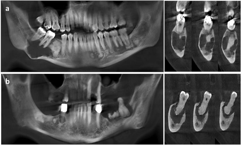
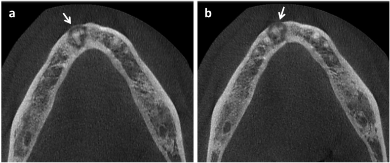
The study group included CBCT images of 142 patients (130 females (91.5%) and 12 males (8.5%)) with a mean age of 46.97 ± 10.57 years. The mandible was involved in almost all cases (99.3%). The most common subtype was florid COD (51.4%) and lesions with hyperdense internal density (81.7%) were more commonly observed. Cortical thinning (78.2%) was a prominent feature. The frequency of root resorption in periapical COD cases (57.1%) was observed to be significantly higher (p < 0.05). All hypercementosis cases were associated with florid subtype (p < 0.05). In a minority of cases (6.3%), the lesions were associated with bone cysts and osteomyelitis.
CBCT images clearly demonstrated the effect of COD lesions on surrounding structures. CBCT is an appropriate imaging modality for the diagnosis and follow-up of COD which is the most common fibro-osseous lesion in clinical practice.
背景/目的:放射学检查在骨化纤维瘤病(COD)的诊断和随访中不可或缺。本回顾性研究的目的是描述一系列COD病例,确定COD亚型的发生率,并研究与亚型相关的人口统计学和放射学特征。
本研究纳入了诊断为COD的患者的锥形束计算机断层扫描(CBCT)图像/报告。收集的数据包括年龄、性别、COD亚型、COD位置和受累区域的信息。还收集了有关内部密度、对周围结构的影响以及伴随病变的存在情况的信息。对获得的数据进行统计学评估。
研究组包括142例患者的CBCT图像(130例女性(91.5%)和12例男性(8.5%)),平均年龄为46.97±10.57岁。几乎所有病例(99.3%)下颌骨均受累。最常见的亚型是弥漫性COD(51.4%),内部密度为高密度的病变(81.7%)更常见。皮质变薄(78.2%)是一个突出特征。根尖周COD病例中牙根吸收的发生率(57.1%)显著更高(p<0.05)。所有牙骨质增生病例均与弥漫性亚型相关(p<0.05)。在少数病例(6.3%)中,病变与骨囊肿和骨髓炎有关。
CBCT图像清楚地显示了COD病变对周围结构的影响。CBCT是临床实践中最常见的纤维-骨病变COD诊断和随访的合适影像学检查方法。