• 文献检索
  • 文档翻译
  • 深度研究
  • 学术资讯
  • Suppr Zotero 插件Zotero 插件
  • 邀请有礼
  • 套餐&价格
  • 历史记录
应用&插件
Suppr Zotero 插件Zotero 插件浏览器插件Mac 客户端Windows 客户端微信小程序
定价
高级版会员购买积分包购买API积分包
服务
文献检索文档翻译深度研究API 文档MCP 服务
关于我们
关于 Suppr公司介绍联系我们用户协议隐私条款
关注我们

Suppr 超能文献

核心技术专利:CN118964589B侵权必究
粤ICP备2023148730 号-1Suppr @ 2026

文献检索

告别复杂PubMed语法,用中文像聊天一样搜索,搜遍4000万医学文献。AI智能推荐,让科研检索更轻松。

立即免费搜索

文件翻译

保留排版,准确专业,支持PDF/Word/PPT等文件格式,支持 12+语言互译。

免费翻译文档

深度研究

AI帮你快速写综述,25分钟生成高质量综述,智能提取关键信息,辅助科研写作。

立即免费体验

利用 CT 胸部图像的放射组学特征对 2019 年冠状病毒病(COVID-19)和其他肺炎进行诊断分类。

Diagnostic classification of coronavirus disease 2019 (COVID-19) and other pneumonias using radiomics features in CT chest images.

机构信息

Department of Medical Imaging, Guangdong Second Provincial General Hospital, Guangzhou, 510317, People's Republic of China.

Radiology Department, Xiao Chang First People's Hospital, Hubei, People's Republic of China.

出版信息

Sci Rep. 2021 Sep 9;11(1):17885. doi: 10.1038/s41598-021-97497-9.

DOI:10.1038/s41598-021-97497-9
PMID:34504246
原文链接:https://pmc.ncbi.nlm.nih.gov/articles/PMC8429652/
Abstract

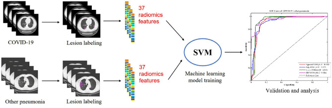
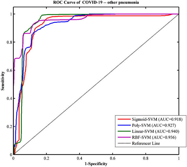
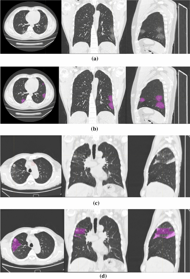
We propose a classification method using the radiomics features of CT chest images to identify patients with coronavirus disease 2019 (COVID-19) and other pneumonias. The chest CT images of two groups of participants (90 COVID-19 patients who were confirmed as positive by nucleic acid test of RT-PCR and 90 other pneumonias patients) were collected, and the two groups of data were manually drawn to outline the region of interest (ROI) of pneumonias. The radiomics method was used to extract textural features and histogram features of the ROI and obtain a radiomics features vector from each sample. Then, we divided the data into two independent radiomic cohorts for training (70 COVID-19 patients and 70 other pneumonias patients), and validation (20 COVID-19 patients and 20 other pneumonias patients) by using support vector machine (SVM). This model used 20 rounds of tenfold cross-validation for training. Finally, single-shot testing of the final model was performed on the independent validation cohort. In the COVID-19 patients, correlation analysis (multiple comparison correction-Bonferroni correction, P < 0.05/7) was also conducted to determine whether the textural and histogram features were correlated with the laboratory test index of blood, i.e., blood oxygen, white blood cell, lymphocytes, neutrophils, C-reactive protein, hypersensitive C-reactive protein, and erythrocyte sedimentation rate. The final model showed good discrimination on the independent validation cohort, with an accuracy of 89.83%, sensitivity of 94.22%, specificity of 85.44%, and AUC of 0.940. This proved that the radiomics features were highly distinguishable, and this SVM model can effectively identify and diagnose patients with COVID-19 and other pneumonias. The correlation analysis results showed that some textural features were positively correlated with WBC, and NE, and also negatively related to SPO2H and NE. Our results showed that radiomic features can classify COVID-19 patients and other pneumonias patients. The SVM model can achieve an excellent diagnosis of COVID-19.

摘要

我们提出了一种利用 CT 胸部图像的放射组学特征来识别 2019 年冠状病毒病(COVID-19)和其他肺炎患者的分类方法。收集了两组参与者的胸部 CT 图像(90 例经 RT-PCR 核酸检测确诊为阳性的 COVID-19 患者和 90 例其他肺炎患者),并手动勾勒出肺炎的感兴趣区域(ROI)。利用放射组学方法提取 ROI 的纹理特征和直方图特征,并从每个样本中获取放射组学特征向量。然后,我们将数据分为两个独立的放射组学队列进行训练(70 例 COVID-19 患者和 70 例其他肺炎患者)和验证(20 例 COVID-19 患者和 20 例其他肺炎患者),并采用支持向量机(SVM)进行分析。该模型使用 20 轮 10 折交叉验证进行训练。最后,在独立验证队列上对最终模型进行单次测试。在 COVID-19 患者中,还进行了相关性分析(多重比较校正- Bonferroni 校正,P<0.05/7),以确定纹理和直方图特征是否与血液的实验室检查指标(如血氧、白细胞、淋巴细胞、中性粒细胞、C 反应蛋白、超敏 C 反应蛋白和红细胞沉降率)相关。最终模型在独立验证队列上具有良好的判别能力,准确率为 89.83%,敏感度为 94.22%,特异度为 85.44%,AUC 为 0.940。这证明放射组学特征具有高度的可区分性,该 SVM 模型可以有效地识别和诊断 COVID-19 和其他肺炎患者。相关性分析结果表明,一些纹理特征与 WBC 和 NE 呈正相关,与 SPO2H 和 NE 呈负相关。我们的结果表明,放射组学特征可以对 COVID-19 患者和其他肺炎患者进行分类。SVM 模型可以实现对 COVID-19 的出色诊断。

https://cdn.ncbi.nlm.nih.gov/pmc/blobs/98f8/8429652/b96e57d63b6b/41598_2021_97497_Fig3_HTML.jpg
https://cdn.ncbi.nlm.nih.gov/pmc/blobs/98f8/8429652/1d5e0877e20c/41598_2021_97497_Fig1_HTML.jpg
https://cdn.ncbi.nlm.nih.gov/pmc/blobs/98f8/8429652/e4cb1e73a7f8/41598_2021_97497_Fig2_HTML.jpg
https://cdn.ncbi.nlm.nih.gov/pmc/blobs/98f8/8429652/b96e57d63b6b/41598_2021_97497_Fig3_HTML.jpg
https://cdn.ncbi.nlm.nih.gov/pmc/blobs/98f8/8429652/1d5e0877e20c/41598_2021_97497_Fig1_HTML.jpg
https://cdn.ncbi.nlm.nih.gov/pmc/blobs/98f8/8429652/e4cb1e73a7f8/41598_2021_97497_Fig2_HTML.jpg
https://cdn.ncbi.nlm.nih.gov/pmc/blobs/98f8/8429652/b96e57d63b6b/41598_2021_97497_Fig3_HTML.jpg

相似文献

1
Diagnostic classification of coronavirus disease 2019 (COVID-19) and other pneumonias using radiomics features in CT chest images.利用 CT 胸部图像的放射组学特征对 2019 年冠状病毒病(COVID-19)和其他肺炎进行诊断分类。
Sci Rep. 2021 Sep 9;11(1):17885. doi: 10.1038/s41598-021-97497-9.
2
Machine learning-based CT radiomics model distinguishes COVID-19 from non-COVID-19 pneumonia.基于机器学习的 CT 放射组学模型可区分 COVID-19 与非 COVID-19 肺炎。
BMC Infect Dis. 2021 Sep 8;21(1):931. doi: 10.1186/s12879-021-06614-6.
3
A COVID-19 risk score combining chest CT radiomics and clinical characteristics to differentiate COVID-19 pneumonia from other viral pneumonias.一种联合胸部 CT 放射组学和临床特征的 COVID-19 风险评分,用于区分 COVID-19 肺炎与其他病毒性肺炎。
Aging (Albany NY). 2021 Mar 13;13(7):9186-9224. doi: 10.18632/aging.202735.
4
A novel CT-based radiomics in the distinction of severity of coronavirus disease 2019 (COVID-19) pneumonia.一种基于 CT 的新型放射组学在区分 2019 年冠状病毒病(COVID-19)肺炎严重程度中的应用。
BMC Infect Dis. 2021 Jun 25;21(1):608. doi: 10.1186/s12879-021-06331-0.
5
Screening of COVID-19 based on the extracted radiomics features from chest CT images.基于胸部 CT 图像提取的放射组学特征对 COVID-19 进行筛查。
J Xray Sci Technol. 2021;29(2):229-243. doi: 10.3233/XST-200831.
6
CT-based radiomics for predicting the rapid progression of coronavirus disease 2019 (COVID-19) pneumonia lesions.基于 CT 的影像组学预测 2019 冠状病毒病(COVID-19)肺炎病变的快速进展。
Br J Radiol. 2021 Jun 1;94(1122):20201007. doi: 10.1259/bjr.20201007. Epub 2021 Apr 21.
7
CT radiomics facilitates more accurate diagnosis of COVID-19 pneumonia: compared with CO-RADS.CT 放射组学有助于更准确地诊断 COVID-19 肺炎:与 CO-RADS 相比。
J Transl Med. 2021 Jan 7;19(1):29. doi: 10.1186/s12967-020-02692-3.
8
A comparison of clinical, laboratory and chest CT findings of laboratory-confirmed and clinically diagnosed COVID-19 patients at first admission.比较首次入院时经实验室确诊和临床诊断的 COVID-19 患者的临床、实验室和胸部 CT 表现。
Diagn Interv Radiol. 2021 May;27(3):336-343. doi: 10.5152/dir.2020.20270.
9
CT radiomic models to distinguish COVID-19 pneumonia from other interstitial pneumonias.CT 放射组学模型鉴别 COVID-19 肺炎与其他间质性肺炎。
Radiol Med. 2021 Aug;126(8):1037-1043. doi: 10.1007/s11547-021-01370-8. Epub 2021 May 27.
10
Clinical manifestations and radiological features by chest computed tomographic findings of a novel coronavirus disease-19 pneumonia among 92 patients in Japan.日本 92 例新型冠状病毒病肺炎患者的胸部计算机断层扫描结果的临床表现和放射学特征。
J Microbiol Immunol Infect. 2021 Aug;54(4):748-751. doi: 10.1016/j.jmii.2020.07.011. Epub 2020 Aug 20.

引用本文的文献

1
Pulmonary contusion volume and radiomics as predictors of early-onset pneumonia in trauma patients, a retrospective single-centre study.创伤患者肺挫伤体积和影像组学作为早期肺炎预测指标的回顾性单中心研究
Eur J Trauma Emerg Surg. 2025 Aug 22;51(1):275. doi: 10.1007/s00068-025-02952-1.
2
Multivariable Risk Modelling and Survival Analysis with Machine Learning in SARS-CoV-2 Infection.SARS-CoV-2感染中基于机器学习的多变量风险建模与生存分析
J Clin Med. 2023 Nov 18;12(22):7164. doi: 10.3390/jcm12227164.
3
A Novel COVID-19 Diagnosis Approach Utilizing a Comprehensive Set of Diagnostic Information (CSDI).

本文引用的文献

1
CT radiomics facilitates more accurate diagnosis of COVID-19 pneumonia: compared with CO-RADS.CT 放射组学有助于更准确地诊断 COVID-19 肺炎:与 CO-RADS 相比。
J Transl Med. 2021 Jan 7;19(1):29. doi: 10.1186/s12967-020-02692-3.
2
A Radiomics Signature to Quantitatively Analyze COVID-19-Infected Pulmonary Lesions.一种用于定量分析 COVID-19 感染肺部病变的放射组学特征。
Interdiscip Sci. 2021 Mar;13(1):61-72. doi: 10.1007/s12539-020-00410-7. Epub 2021 Jan 7.
3
Development and Validation of an Automated Radiomic CT Signature for Detecting COVID-19.
一种利用综合诊断信息集(CSDI)的新型新冠病毒诊断方法。
J Clin Med. 2023 Nov 3;12(21):6912. doi: 10.3390/jcm12216912.
4
Immunohematologic Biomarkers in COVID-19: Insights into Pathogenesis, Prognosis, and Prevention.COVID-19中的免疫血液生物标志物:对发病机制、预后和预防的见解
Pathog Immun. 2023 Jun 26;8(1):17-50. doi: 10.20411/pai.v8i1.572. eCollection 2023.
5
Toward the determination of sensitive and reliable whole-lung computed tomography features for robust standard radiomics and delta-radiomics analysis in a nonhuman primate model of coronavirus disease 2019.在2019冠状病毒病的非人灵长类动物模型中,确定敏感且可靠的全肺计算机断层扫描特征,以进行稳健的标准放射组学和增量放射组学分析。
J Med Imaging (Bellingham). 2022 Nov;9(6):066003. doi: 10.1117/1.JMI.9.6.066003. Epub 2022 Dec 8.
6
BND-VGG-19: A deep learning algorithm for COVID-19 identification utilizing X-ray images.BND-VGG-19:一种利用X射线图像识别新冠肺炎的深度学习算法。
Knowl Based Syst. 2022 Dec 22;258:110040. doi: 10.1016/j.knosys.2022.110040. Epub 2022 Oct 21.
7
Challenges of Multiplex Assays for COVID-19 Research: A Machine Learning Perspective.用于 COVID-19 研究的多重分析面临的挑战:机器学习视角。
Methods Mol Biol. 2022;2511:37-50. doi: 10.1007/978-1-0716-2395-4_3.
8
Development and Validation of a Radiomics Nomogram Using Computed Tomography for Differentiating Immune Checkpoint Inhibitor-Related Pneumonitis From Radiation Pneumonitis for Patients With Non-Small Cell Lung Cancer.基于 CT 影像的放射组学列线图模型构建及其在免疫检查点抑制剂相关性肺炎与放射性肺炎鉴别诊断中的价值:一项针对非小细胞肺癌患者的研究
Front Immunol. 2022 Apr 26;13:870842. doi: 10.3389/fimmu.2022.870842. eCollection 2022.
9
Interactions of Co, Cu, and non-metal phthalocyanines with external structures of SARS-CoV-2 using docking and molecular dynamics.利用对接和分子动力学研究 Co、Cu 和非金属酞菁与 SARS-CoV-2 外部结构的相互作用。
Sci Rep. 2022 Feb 28;12(1):3316. doi: 10.1038/s41598-022-07396-w.
10
A review on advances in F-FDG PET/CT radiomics standardisation and application in lung disease management.18F-氟代脱氧葡萄糖正电子发射断层显像/计算机断层扫描(18F-FDG PET/CT)影像组学在肺部疾病管理中的标准化进展与应用综述
Insights Imaging. 2022 Feb 5;13(1):22. doi: 10.1186/s13244-021-01153-9.
用于检测新型冠状病毒肺炎的自动化影像组学CT特征的开发与验证
Diagnostics (Basel). 2020 Dec 30;11(1):41. doi: 10.3390/diagnostics11010041.
4
Hypergraph learning for identification of COVID-19 with CT imaging.基于CT成像的超图学习用于新冠病毒肺炎的识别
Med Image Anal. 2021 Feb;68:101910. doi: 10.1016/j.media.2020.101910. Epub 2020 Nov 26.
5
CT Quantification and Machine-learning Models for Assessment of Disease Severity and Prognosis of COVID-19 Patients.CT 定量和机器学习模型评估 COVID-19 患者的疾病严重程度和预后。
Acad Radiol. 2020 Dec;27(12):1665-1678. doi: 10.1016/j.acra.2020.09.004. Epub 2020 Sep 21.
6
Modified SEIR and AI prediction of the epidemics trend of COVID-19 in China under public health interventions.公共卫生干预下中国新冠疫情趋势的改进型SEIR模型及人工智能预测
J Thorac Dis. 2020 Mar;12(3):165-174. doi: 10.21037/jtd.2020.02.64.
7
Which lessons shall we learn from the 2019 novel coronavirus outbreak?我们应从2019年新型冠状病毒疫情中学到哪些教训?
Ann Transl Med. 2020 Feb;8(3):48. doi: 10.21037/atm.2020.02.06.
8
Time Course of Lung Changes at Chest CT during Recovery from Coronavirus Disease 2019 (COVID-19).新冠肺炎(COVID-19)康复过程中胸部 CT 肺部变化的时间进程。
Radiology. 2020 Jun;295(3):715-721. doi: 10.1148/radiol.2020200370. Epub 2020 Feb 13.
9
Chest CT for Typical Coronavirus Disease 2019 (COVID-19) Pneumonia: Relationship to Negative RT-PCR Testing.胸部 CT 与典型 2019 冠状病毒病(COVID-19)肺炎:与阴性 RT-PCR 检测的关系。
Radiology. 2020 Aug;296(2):E41-E45. doi: 10.1148/radiol.2020200343. Epub 2020 Feb 12.
10
Clinical Characteristics of 138 Hospitalized Patients With 2019 Novel Coronavirus-Infected Pneumonia in Wuhan, China.《武汉 2019 年新型冠状病毒感染的肺炎 138 例住院患者临床特征分析》
JAMA. 2020 Mar 17;323(11):1061-1069. doi: 10.1001/jama.2020.1585.