Department of Radiology, Hainan General Hospital (Hainan Affiliated Hospital of Hainan Medical University), No. 19, Xiuhua St, Xiuying Dic, Haikou, Hainan, 570311, People's Republic of China.
Deepwise AI Lab, Deepwise Inc., No. 8 Haidian avenue, Sinosteel International Plaza, Beijing, 100080, China.
BMC Infect Dis. 2021 Sep 8;21(1):931. doi: 10.1186/s12879-021-06614-6.
To develop a machine learning-based CT radiomics model is critical for the accurate diagnosis of the rapid spreading coronavirus disease 2019 (COVID-19).
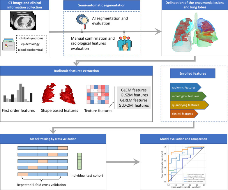
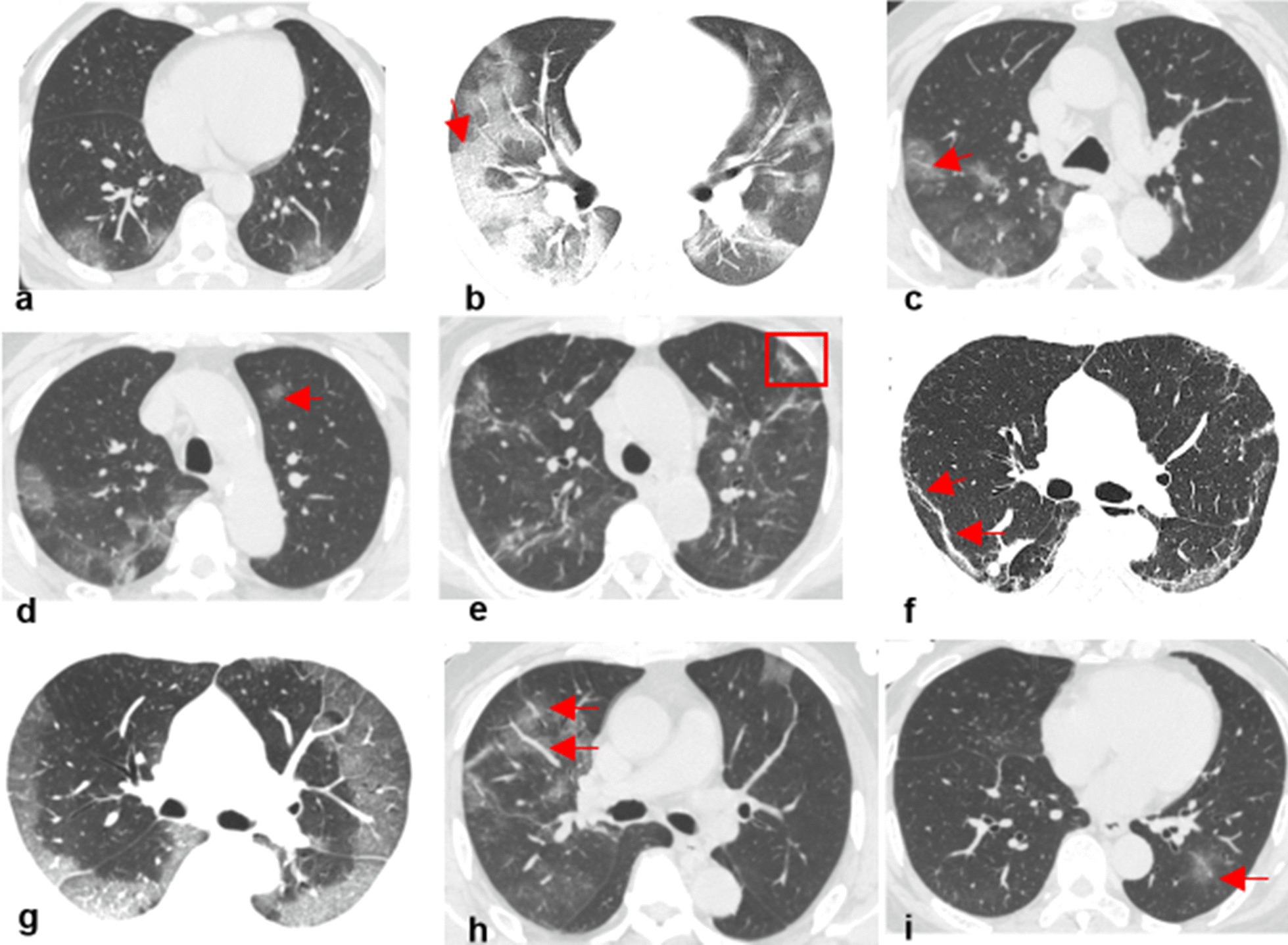
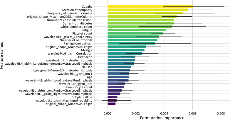
In this retrospective study, a total of 326 chest CT exams from 134 patients (63 confirmed COVID-19 patients and 71 non-COVID-19 patients) were collected from January 20 to February 8, 2020. A semi-automatic segmentation procedure was used to delineate the volume of interest (VOI), and radiomic features were extracted. The Support Vector Machine (SVM) model was built on the combination of 4 groups of features, including radiomic features, traditional radiological features, quantifying features, and clinical features. By repeating cross-validation procedure, the performance on the time-independent testing cohort was evaluated by the area under the receiver operating characteristic curve (AUC), accuracy, sensitivity, and specificity.
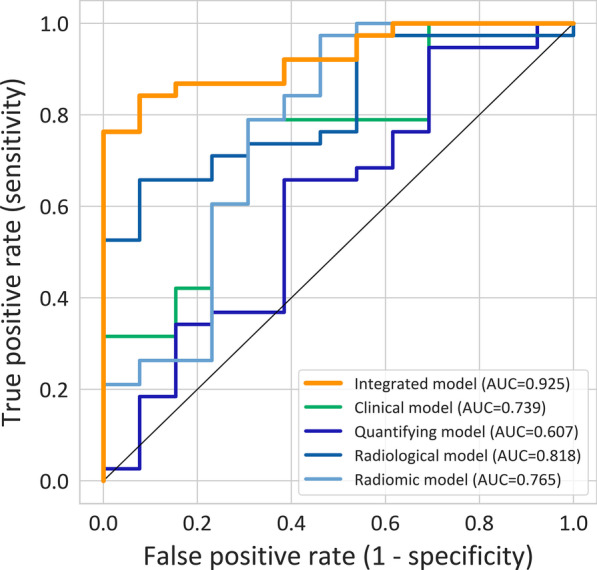
For the SVM model built on the combination of 4 groups of features (integrated model), the per-exam AUC was 0.925 (95% CI 0.856 to 0.994) for differentiating COVID-19 on the testing cohort, and the sensitivity and specificity were 0.816 (95% CI 0.651 to 0.917) and 0.923 (95% CI 0.621 to 0.996), respectively. As for the SVM models built on radiomic features, radiological features, quantifying features, and clinical features, individually, the AUC on the testing cohort reached 0.765, 0.818, 0.607, and 0.739, respectively, significantly lower than the integrated model, except for the radiomic model.
The machine learning-based CT radiomics models may accurately classify COVID-19, helping clinicians and radiologists to identify COVID-19 positive cases.
开发基于机器学习的 CT 放射组学模型对于准确诊断快速传播的 2019 年冠状病毒病(COVID-19)至关重要。
在这项回顾性研究中,共从 2020 年 1 月 20 日至 2 月 8 日收集了 134 名患者(63 名确诊 COVID-19 患者和 71 名非 COVID-19 患者)的 326 次胸部 CT 检查。使用半自动分割程序对感兴趣的容积(VOI)进行描绘,并提取放射组学特征。基于 4 组特征(放射组学特征、传统放射学特征、定量特征和临床特征)的组合构建支持向量机(SVM)模型。通过重复交叉验证程序,使用受试者工作特征曲线下的面积(AUC)、准确性、灵敏度和特异性来评估时间独立测试队列的性能。
对于基于 4 组特征(综合模型)构建的 SVM 模型,在测试队列中区分 COVID-19 的每例检查 AUC 为 0.925(95%CI 0.856 至 0.994),灵敏度和特异性分别为 0.816(95%CI 0.651 至 0.917)和 0.923(95%CI 0.621 至 0.996)。对于基于放射组学特征、放射学特征、定量特征和临床特征分别构建的 SVM 模型,在测试队列中的 AUC 分别达到 0.765、0.818、0.607 和 0.739,明显低于综合模型,除了放射组学模型。
基于机器学习的 CT 放射组学模型可以准确分类 COVID-19,帮助临床医生和放射科医生识别 COVID-19 阳性病例。