• 文献检索
  • 文档翻译
  • 深度研究
  • 学术资讯
  • Suppr Zotero 插件Zotero 插件
  • 邀请有礼
  • 套餐&价格
  • 历史记录
应用&插件
Suppr Zotero 插件Zotero 插件浏览器插件Mac 客户端Windows 客户端微信小程序
定价
高级版会员购买积分包购买API积分包
服务
文献检索文档翻译深度研究API 文档MCP 服务
关于我们
关于 Suppr公司介绍联系我们用户协议隐私条款
关注我们

Suppr 超能文献

核心技术专利:CN118964589B侵权必究
粤ICP备2023148730 号-1Suppr @ 2026

文献检索

告别复杂PubMed语法,用中文像聊天一样搜索,搜遍4000万医学文献。AI智能推荐,让科研检索更轻松。

立即免费搜索

文件翻译

保留排版,准确专业,支持PDF/Word/PPT等文件格式,支持 12+语言互译。

免费翻译文档

深度研究

AI帮你快速写综述,25分钟生成高质量综述,智能提取关键信息,辅助科研写作。

立即免费体验

用于检测新型冠状病毒肺炎的自动化影像组学CT特征的开发与验证

Development and Validation of an Automated Radiomic CT Signature for Detecting COVID-19.

作者信息

Guiot Julien, Vaidyanathan Akshayaa, Deprez Louis, Zerka Fadila, Danthine Denis, Frix Anne-Noëlle, Thys Marie, Henket Monique, Canivet Gregory, Mathieu Stephane, Eftaxia Evanthia, Lambin Philippe, Tsoutzidis Nathan, Miraglio Benjamin, Walsh Sean, Moutschen Michel, Louis Renaud, Meunier Paul, Vos Wim, Leijenaar Ralph T H, Lovinfosse Pierre

机构信息

Department of Pneumology, University Hospital of Liège, 4020 Liège, Belgium.

Research and Development, Oncoradiomics SA, 4000 Liège, Belgium.

出版信息

Diagnostics (Basel). 2020 Dec 30;11(1):41. doi: 10.3390/diagnostics11010041.

DOI:10.3390/diagnostics11010041
PMID:33396587
原文链接:https://pmc.ncbi.nlm.nih.gov/articles/PMC7823620/
Abstract

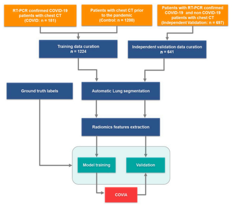
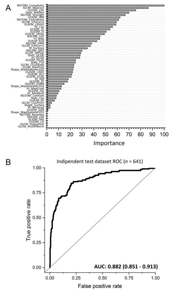
The coronavirus disease 2019 (COVID-19) outbreak has reached pandemic status. Drastic measures of social distancing are enforced in society and healthcare systems are being pushed to and beyond their limits. To help in the fight against this threat on human health, a fully automated AI framework was developed to extract radiomics features from volumetric chest computed tomography (CT) exams. The detection model was developed on a dataset of 1381 patients (181 COVID-19 patients plus 1200 non COVID control patients). A second, independent dataset of 197 RT-PCR confirmed COVID-19 patients and 500 control patients was used to assess the performance of the model. Diagnostic performance was assessed by the area under the receiver operating characteristic curve (AUC). The model had an AUC of 0.882 (95% CI: 0.851-0.913) in the independent test dataset (641 patients). The optimal decision threshold, considering the cost of false negatives twice as high as the cost of false positives, resulted in an accuracy of 85.18%, a sensitivity of 69.52%, a specificity of 91.63%, a negative predictive value (NPV) of 94.46% and a positive predictive value (PPV) of 59.44%. Benchmarked against RT-PCR confirmed cases of COVID-19, our AI framework can accurately differentiate COVID-19 from routine clinical conditions in a fully automated fashion. Thus, providing rapid accurate diagnosis in patients suspected of COVID-19 infection, facilitating the timely implementation of isolation procedures and early intervention.

摘要

2019冠状病毒病(COVID-19)疫情已达到大流行状态。社会上实施了严格的社交距离措施,医疗系统面临着巨大压力,甚至超出了其极限。为了帮助应对这一人类健康威胁,开发了一个全自动人工智能框架,用于从胸部容积计算机断层扫描(CT)检查中提取放射组学特征。检测模型是基于一个包含1381名患者的数据集开发的(181名COVID-19患者加上1200名非COVID对照患者)。另一个由197名经逆转录聚合酶链反应(RT-PCR)确诊的COVID-19患者和500名对照患者组成的独立数据集用于评估该模型的性能。通过受试者操作特征曲线(AUC)下的面积评估诊断性能。该模型在独立测试数据集(641名患者)中的AUC为0.882(95%CI:0.851-0.913)。考虑到假阴性成本是假阳性成本的两倍,最佳决策阈值下的准确率为85.18%,灵敏度为69.52%,特异性为91.63%,阴性预测值(NPV)为94.46%。,阳性预测值(PPV)为59.44%。与RT-PCR确诊的COVID-19病例相比,我们的人工智能框架可以以全自动方式准确区分COVID-19与常规临床情况。因此,能够对疑似COVID-19感染的患者进行快速准确的诊断,便于及时实施隔离程序和早期干预。

https://cdn.ncbi.nlm.nih.gov/pmc/blobs/a46c/7823620/45d7ea5852ec/diagnostics-11-00041-g006.jpg
https://cdn.ncbi.nlm.nih.gov/pmc/blobs/a46c/7823620/92b79647ee75/diagnostics-11-00041-g001.jpg
https://cdn.ncbi.nlm.nih.gov/pmc/blobs/a46c/7823620/0bde26ffb061/diagnostics-11-00041-g002.jpg
https://cdn.ncbi.nlm.nih.gov/pmc/blobs/a46c/7823620/e3ff0f0e8eb6/diagnostics-11-00041-g003.jpg
https://cdn.ncbi.nlm.nih.gov/pmc/blobs/a46c/7823620/5e114c2f5ae1/diagnostics-11-00041-g004.jpg
https://cdn.ncbi.nlm.nih.gov/pmc/blobs/a46c/7823620/c6d5ad8eadb6/diagnostics-11-00041-g005.jpg
https://cdn.ncbi.nlm.nih.gov/pmc/blobs/a46c/7823620/45d7ea5852ec/diagnostics-11-00041-g006.jpg

相似文献

1
Development and Validation of an Automated Radiomic CT Signature for Detecting COVID-19.用于检测新型冠状病毒肺炎的自动化影像组学CT特征的开发与验证
Diagnostics (Basel). 2020 Dec 30;11(1):41. doi: 10.3390/diagnostics11010041.
2
Machine learning-based CT radiomics model distinguishes COVID-19 from non-COVID-19 pneumonia.基于机器学习的 CT 放射组学模型可区分 COVID-19 与非 COVID-19 肺炎。
BMC Infect Dis. 2021 Sep 8;21(1):931. doi: 10.1186/s12879-021-06614-6.
3
Machine learning-based prognostic modeling using clinical data and quantitative radiomic features from chest CT images in COVID-19 patients.基于机器学习的 COVID-19 患者胸部 CT 图像临床数据和定量放射组学特征的预后建模。
Comput Biol Med. 2021 May;132:104304. doi: 10.1016/j.compbiomed.2021.104304. Epub 2021 Mar 3.
4
Using Artificial Intelligence to Detect COVID-19 and Community-acquired Pneumonia Based on Pulmonary CT: Evaluation of the Diagnostic Accuracy.基于肺部 CT 的人工智能检测 COVID-19 和社区获得性肺炎:诊断准确性评估。
Radiology. 2020 Aug;296(2):E65-E71. doi: 10.1148/radiol.2020200905. Epub 2020 Mar 19.
5
CT radiomics facilitates more accurate diagnosis of COVID-19 pneumonia: compared with CO-RADS.CT 放射组学有助于更准确地诊断 COVID-19 肺炎:与 CO-RADS 相比。
J Transl Med. 2021 Jan 7;19(1):29. doi: 10.1186/s12967-020-02692-3.
6
A model based on CT radiomic features for predicting RT-PCR becoming negative in coronavirus disease 2019 (COVID-19) patients.基于 CT 影像组学特征的模型预测 2019 冠状病毒病(COVID-19)患者 RT-PCR 转为阴性。
BMC Med Imaging. 2020 Oct 20;20(1):118. doi: 10.1186/s12880-020-00521-z.
7
Chest CT in patients with a moderate or high pretest probability of COVID-19 and negative swab.胸部 CT 检查在中度或高度疑似 COVID-19 且咽拭子检测阴性的患者中的应用。
Radiol Med. 2020 Dec;125(12):1260-1270. doi: 10.1007/s11547-020-01269-w. Epub 2020 Aug 29.
8
Non-contrast CT-based radiomic signature for screening thoracic aortic dissections: a multicenter study.基于非增强 CT 的放射组学特征用于筛查胸主动脉夹层:一项多中心研究。
Eur Radiol. 2021 Sep;31(9):7067-7076. doi: 10.1007/s00330-021-07768-2. Epub 2021 Mar 23.
9
Dynamic 3D radiomics analysis using artificial intelligence to assess the stage of COVID-19 on CT images.利用人工智能进行动态 3D 放射组学分析,以评估 CT 图像上 COVID-19 的分期。
Eur Radiol. 2022 Jul;32(7):4760-4770. doi: 10.1007/s00330-021-08533-1. Epub 2022 Jan 29.
10
AI detection of mild COVID-19 pneumonia from chest CT scans.人工智能检测胸部 CT 扫描中的轻症 COVID-19 肺炎。
Eur Radiol. 2021 Sep;31(9):7192-7201. doi: 10.1007/s00330-021-07797-x. Epub 2021 Mar 18.

引用本文的文献

1
Central artery pulse pressure, not central arterial stiffness impact on all-cause mortality in patients with viral pneumonia infection.中央动脉脉压,而非中央动脉僵硬度,影响病毒性肺炎感染患者的全因死亡率。
BMC Infect Dis. 2024 Oct 21;24(1):1183. doi: 10.1186/s12879-024-10091-y.
2
Long Term Evaluation of Quantitative Cumulative Irradiation in Patients Suffering from ILDs.间质性肺疾病患者定量累积照射的长期评估
Diagnostics (Basel). 2024 Sep 26;14(19):2136. doi: 10.3390/diagnostics14192136.
3
Using artificial intelligence and predictive modelling to enable learning healthcare systems (LHS) for pandemic preparedness.

本文引用的文献

1
How Might AI and Chest Imaging Help Unravel COVID-19's Mysteries?人工智能和胸部成像如何助力解开新冠病毒的谜团?
Radiol Artif Intell. 2020 May 6;2(3):e200053. doi: 10.1148/ryai.2020200053. eCollection 2020 May.
2
Deep Learning Enables Accurate Diagnosis of Novel Coronavirus (COVID-19) With CT Images.深度学习利用 CT 图像准确诊断新型冠状病毒(COVID-19)。
IEEE/ACM Trans Comput Biol Bioinform. 2021 Nov-Dec;18(6):2775-2780. doi: 10.1109/TCBB.2021.3065361. Epub 2021 Dec 8.
3
A deep learning algorithm using CT images to screen for Corona virus disease (COVID-19).
利用人工智能和预测模型构建用于大流行防范的学习型医疗系统(LHS)。
Comput Struct Biotechnol J. 2024 May 17;24:412-419. doi: 10.1016/j.csbj.2024.05.014. eCollection 2024 Dec.
4
Comparing Visual and Software-Based Quantitative Assessment Scores of Lungs' Parenchymal Involvement Quantification in COVID-19 Patients.比较COVID-19患者肺部实质受累量化的视觉和基于软件的定量评估分数
Diagnostics (Basel). 2024 May 8;14(10):985. doi: 10.3390/diagnostics14100985.
5
Does Facemask Impact Diagnostic During Pulmonary Auscultation?口罩对肺部听诊诊断有影响吗?
IFAC Pap OnLine. 2021;54(15):192-197. doi: 10.1016/j.ifacol.2021.10.254. Epub 2021 Nov 2.
6
Utility of bronchoalveolar lavage for COVID-19: a perspective from the Dragon consortium.支气管肺泡灌洗在新型冠状病毒肺炎中的应用:来自“龙”联盟的观点
Front Med (Lausanne). 2024 Feb 2;11:1259570. doi: 10.3389/fmed.2024.1259570. eCollection 2024.
7
Multivariable Risk Modelling and Survival Analysis with Machine Learning in SARS-CoV-2 Infection.SARS-CoV-2感染中基于机器学习的多变量风险建模与生存分析
J Clin Med. 2023 Nov 18;12(22):7164. doi: 10.3390/jcm12227164.
8
A Systematic Review on Deep Structured Learning for COVID-19 Screening Using Chest CT from 2020 to 2022.2020年至2022年基于胸部CT的COVID-19筛查深度结构化学习系统综述
Healthcare (Basel). 2023 Aug 24;11(17):2388. doi: 10.3390/healthcare11172388.
9
Quantitative Evaluation of COVID-19 Pneumonia CT Using AI Analysis-Feasibility and Differentiation from Other Common Pneumonia Forms.使用人工智能分析对新型冠状病毒肺炎CT进行定量评估——可行性及与其他常见肺炎形式的鉴别
Diagnostics (Basel). 2023 Jun 20;13(12):2129. doi: 10.3390/diagnostics13122129.
10
Multi-center Integrating Radiomics, Structured Reports, and Machine Learning Algorithms for Assisted Classification of COVID-19 in Lung Computed Tomography.多中心整合影像组学、结构化报告和机器学习算法用于肺部计算机断层扫描中新型冠状病毒肺炎的辅助分类
J Med Biol Eng. 2023;43(2):156-162. doi: 10.1007/s40846-023-00781-4. Epub 2023 Mar 7.
利用 CT 图像进行冠状病毒病(COVID-19)筛查的深度学习算法。
Eur Radiol. 2021 Aug;31(8):6096-6104. doi: 10.1007/s00330-021-07715-1. Epub 2021 Feb 24.
4
Deep learning-based model for detecting 2019 novel coronavirus pneumonia on high-resolution computed tomography.基于深度学习的高分辨率计算机断层扫描 2019 年新型冠状病毒肺炎检测模型。
Sci Rep. 2020 Nov 5;10(1):19196. doi: 10.1038/s41598-020-76282-0.
5
Review on Diagnosis of COVID-19 from Chest CT Images Using Artificial Intelligence.基于人工智能的胸部 CT 图像新冠肺炎诊断研究进展
Comput Math Methods Med. 2020 Sep 26;2020:9756518. doi: 10.1155/2020/9756518. eCollection 2020.
6
Discrimination of pulmonary ground-glass opacity changes in COVID-19 and non-COVID-19 patients using CT radiomics analysis.使用CT影像组学分析鉴别新型冠状病毒肺炎和非新型冠状病毒肺炎患者的肺磨玻璃影变化
Eur J Radiol Open. 2020;7:100271. doi: 10.1016/j.ejro.2020.100271. Epub 2020 Sep 16.
7
Artificial intelligence for the detection of COVID-19 pneumonia on chest CT using multinational datasets.利用多国数据集的人工智能检测胸部 CT 中的 COVID-19 肺炎。
Nat Commun. 2020 Aug 14;11(1):4080. doi: 10.1038/s41467-020-17971-2.
8
Chest CT in COVID-19 pneumonia: A review of current knowledge.胸部 CT 在 COVID-19 肺炎中的应用:现有知识综述。
Diagn Interv Imaging. 2020 Jul-Aug;101(7-8):431-437. doi: 10.1016/j.diii.2020.06.001. Epub 2020 Jun 11.
9
A Novel Machine Learning-derived Radiomic Signature of the Whole Lung Differentiates Stable From Progressive COVID-19 Infection: A Retrospective Cohort Study.一种基于新型机器学习的全肺放射组学特征可区分新冠病毒感染的稳定期与进展期:一项回顾性队列研究。
J Thorac Imaging. 2020 Nov 1;35(6):361-368. doi: 10.1097/RTI.0000000000000544.
10
Artificial intelligence-enabled rapid diagnosis of patients with COVID-19.人工智能助力 COVID-19 患者快速诊断。
Nat Med. 2020 Aug;26(8):1224-1228. doi: 10.1038/s41591-020-0931-3. Epub 2020 May 19.