Department of Orthopaedics, First Affiliated Hospital of Nanjing Medical University, Nanjing, 210029, Jiangsu, China.
J Neuroinflammation. 2021 Sep 12;18(1):196. doi: 10.1186/s12974-021-02268-y.
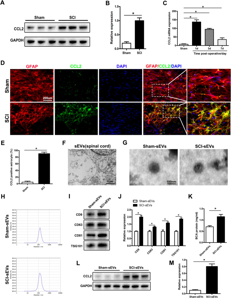
Spinal cord injury (SCI) is a severe traumatic disease which causes high disability and mortality rates. The molecular pathological features after spinal cord injury mainly involve the inflammatory response, microglial and neuronal apoptosis, abnormal proliferation of astrocytes, and the formation of glial scars. However, the microenvironmental changes after spinal cord injury are complex, and the interactions between glial cells and nerve cells remain unclear. Small extracellular vesicles (sEVs) may play a key role in cell communication by transporting RNA, proteins, and bioactive lipids between cells. Few studies have examined the intercellular communication of astrocytes through sEVs after SCI. The inflammatory signal released from astrocytes is known to initiate microglial activation, but its effects on neurons after SCI remain to be further clarified.
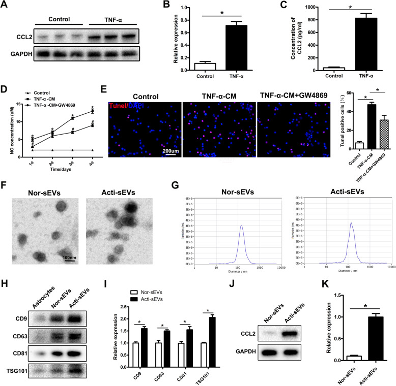
Electron microscopy (TEM), nanoparticle tracking analysis (NTA), and western blotting were applied to characterize sEVs. We examined microglial activation and neuronal apoptosis mediated by astrocyte activation in an experimental model of acute spinal cord injury and in cell culture in vitro.
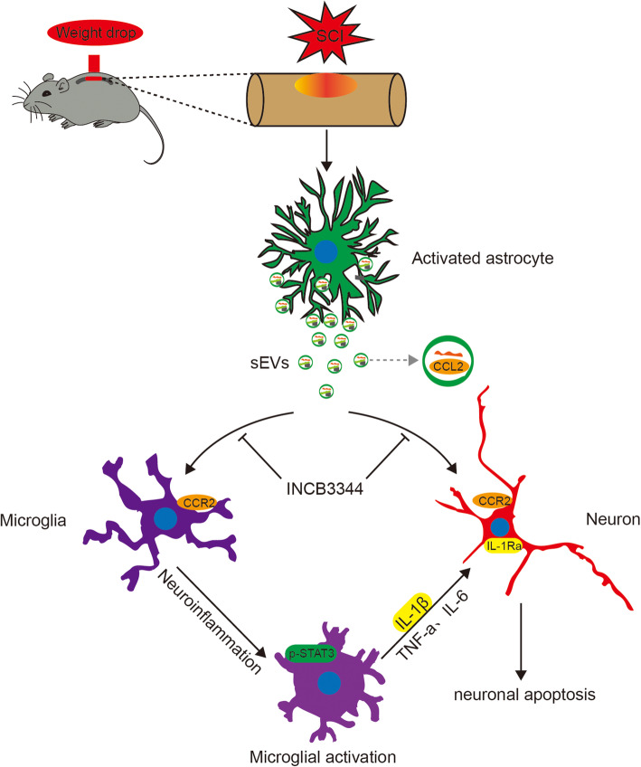
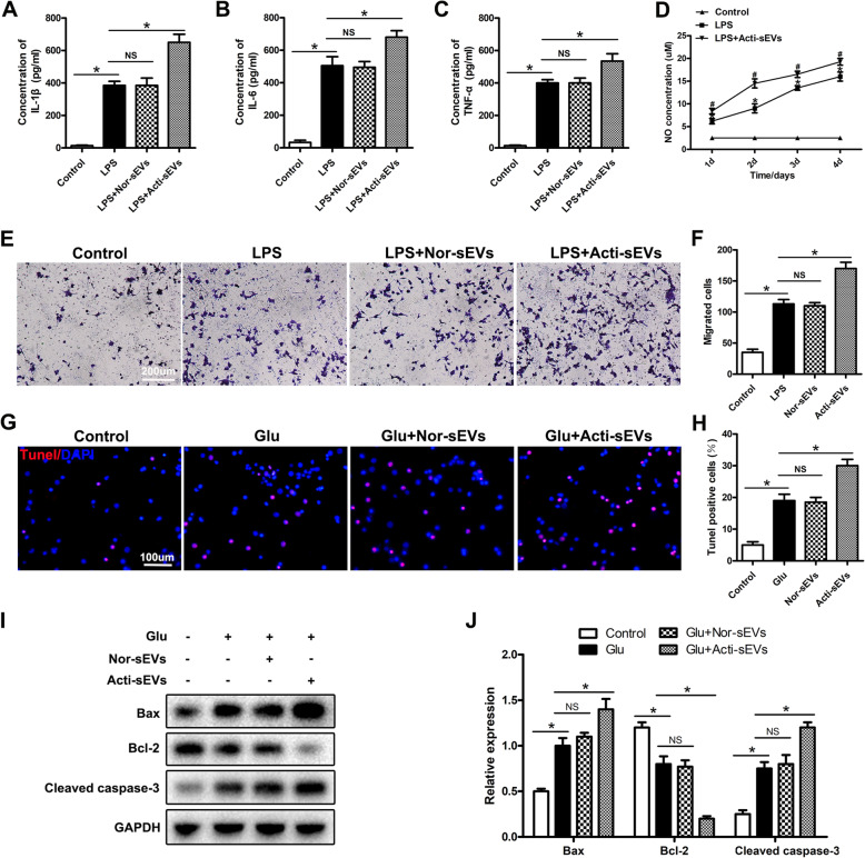
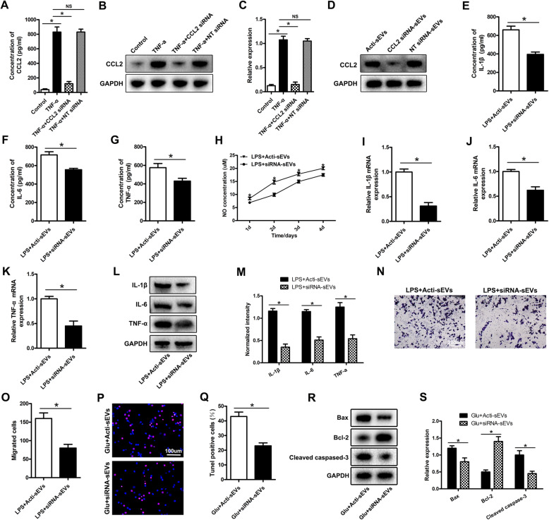
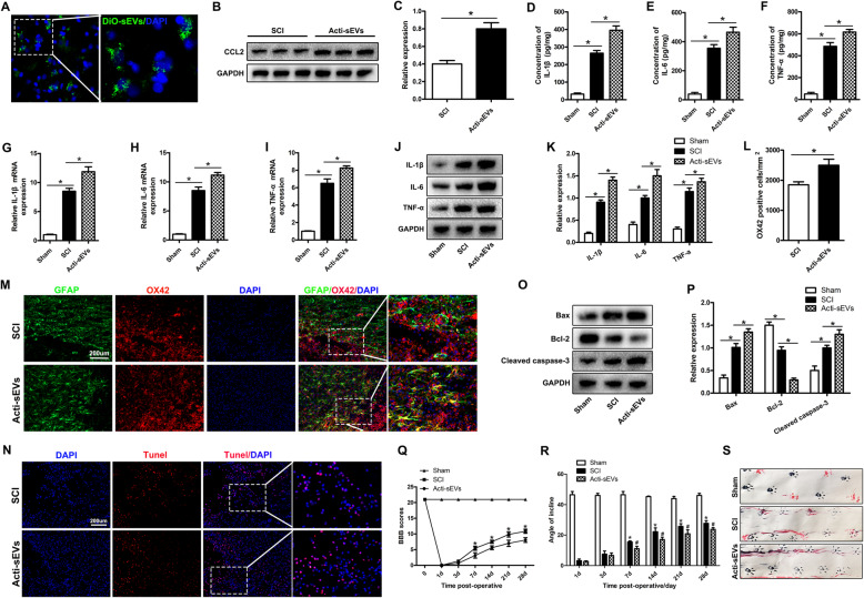
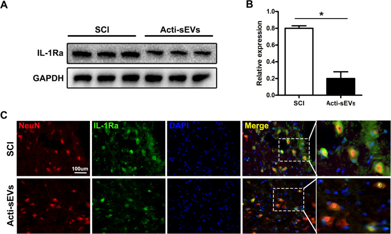
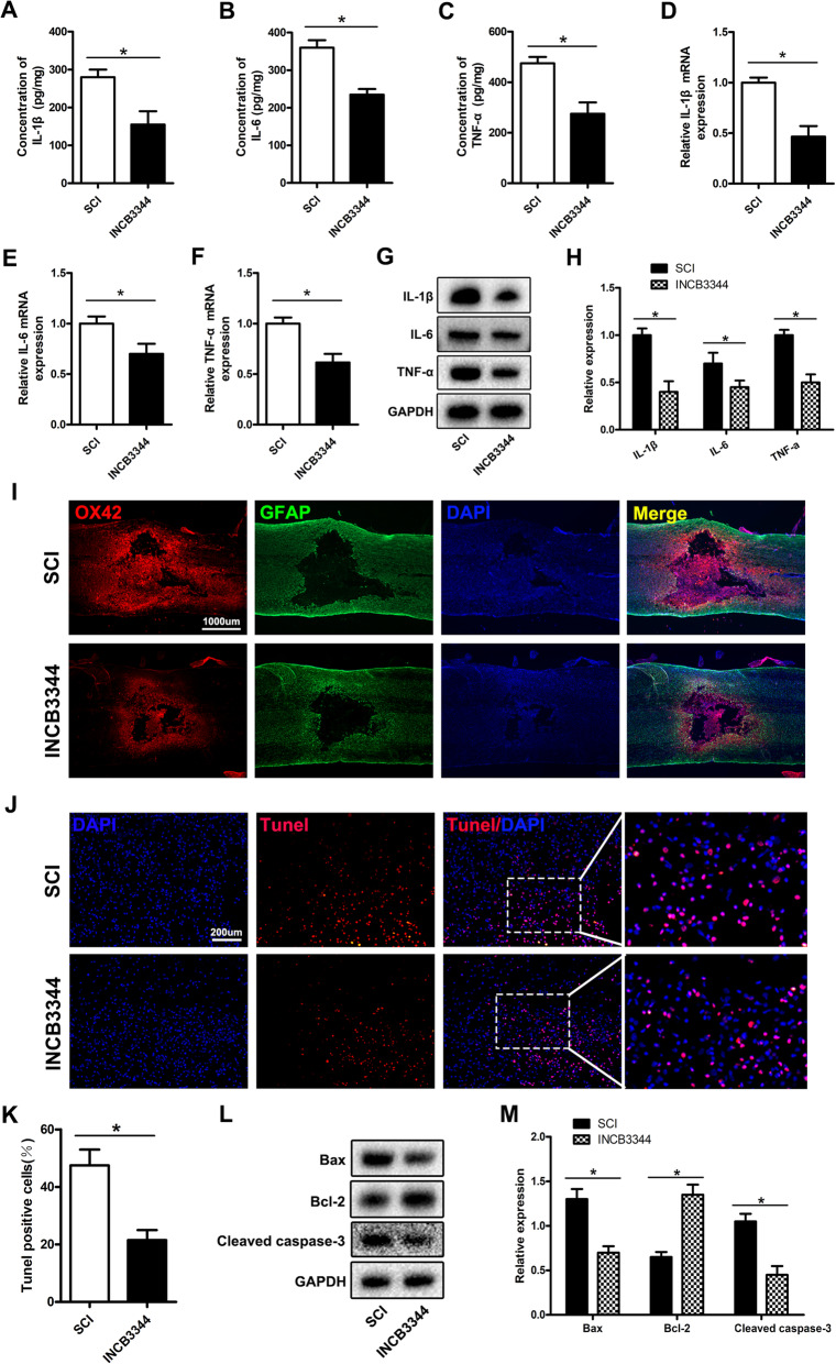
Our results indicated that astrocytes activated after spinal cord injury release CCL2, act on microglia and neuronal cells through the sEV pathway, and promote neuronal apoptosis and microglial activation after binding the CCR2. Subsequently, the activated microglia release IL-1β, which acts on neuronal cells, thereby further aggravating their apoptosis.
This study elucidates that astrocytes interact with microglia and neurons through the sEV pathway after SCI, enriching the mechanism of CCL2 in neuroinflammation and spinal neurodegeneration, and providing a new theoretical basis of CCL2 as a therapeutic target for SCI.
脊髓损伤(SCI)是一种严重的创伤性疾病,导致高残疾率和死亡率。脊髓损伤后的分子病理特征主要涉及炎症反应、小胶质细胞和神经元凋亡、星形胶质细胞异常增殖和胶质瘢痕形成。然而,脊髓损伤后的微环境变化复杂,胶质细胞与神经细胞之间的相互作用尚不清楚。小细胞外囊泡(sEVs)可能通过在细胞间运输 RNA、蛋白质和生物活性脂质在细胞通讯中发挥关键作用。很少有研究探讨过 SCI 后通过 sEVs 进行的星形胶质细胞间的细胞通讯。已知星形胶质细胞释放的炎症信号会引发小胶质细胞激活,但它对 SCI 后神经元的影响仍需进一步阐明。
电子显微镜(TEM)、纳米颗粒跟踪分析(NTA)和 Western blot 用于表征 sEVs。我们在急性脊髓损伤的实验模型和体外细胞培养中研究了星形胶质细胞激活介导的小胶质细胞激活和神经元凋亡。
我们的结果表明,脊髓损伤后激活的星形胶质细胞释放 CCL2,通过 sEV 途径作用于小胶质细胞和神经元细胞,并在与 CCR2 结合后促进神经元凋亡和小胶质细胞激活。随后,激活的小胶质细胞释放出作用于神经元细胞的 IL-1β,从而进一步加重其凋亡。
本研究阐明了 SCI 后星形胶质细胞通过 sEV 途径与小胶质细胞和神经元相互作用,丰富了 CCL2 在神经炎症和脊髓神经退行性变中的作用机制,并为 CCL2 作为 SCI 治疗靶点提供了新的理论依据。