Ha Nguyen Thi Thu, Huong Lai Thu, Trang Dam Thuy, Nhung Luu Hong, Huyen Nguyen Thi, Huy Nguyen Quang, Cuong Do Duy, Luu Vu Dang
Department of Radiology, Hanoi Medical University, Hanoi, Viet Nam.
Bach Mai Radiology Center, Bach Mai hospital, Hanoi, Viet Nam.
Radiol Case Rep. 2021 Sep 5;16(11):3434-3437. doi: 10.1016/j.radcr.2021.08.018. eCollection 2021 Nov.
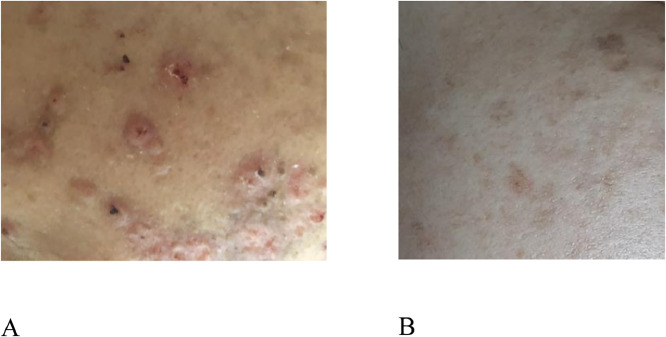
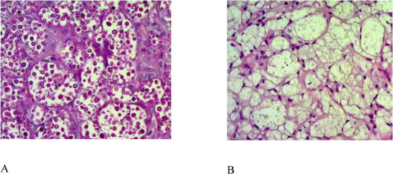
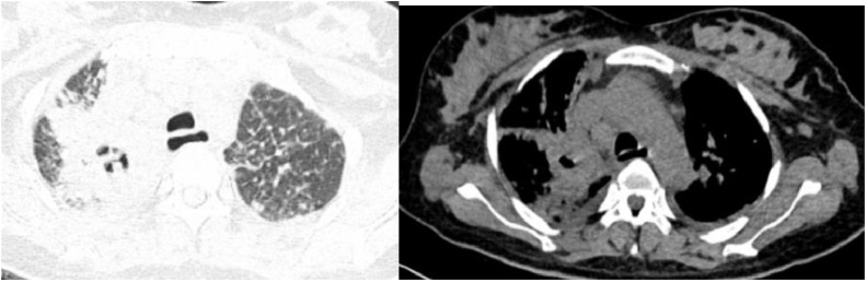
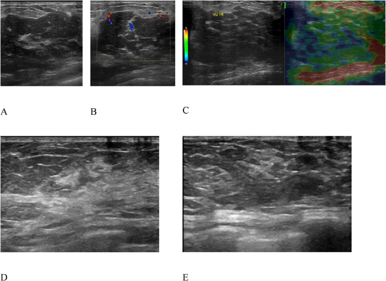
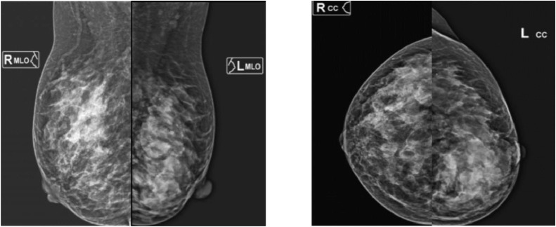
There have been few reports on the imaging characteristics of (C. ) infection of the breast. Herein, we reported the imaging features of C. infection of the breast in a 41-year-old woman with immune thrombocytopenic purpura. Bilateral, diffuse, hyperechoic, and well-defined margin lesions were observed on breast ultrasounds. In addition, a global asymmetry in the left breast, and a focal asymmetry in the right breast were observed on mammograms. Breast fine needle aspiration and biopsy results revealed a C. infection. After 5 months of treatment with oral fluconazole and amphotericin B, the lesion on the right breast disappeared on repeated-breast ultrasounds.
关于乳腺念珠菌感染的影像学特征的报道较少。在此,我们报告了一名患有免疫性血小板减少性紫癜的41岁女性乳腺念珠菌感染的影像学特征。乳腺超声检查发现双侧弥漫性高回声、边界清晰的病变。此外,乳房X线摄影显示左乳整体不对称,右乳局灶性不对称。乳腺细针穿刺活检结果显示为念珠菌感染。口服氟康唑和两性霉素B治疗5个月后,右乳病变在重复乳腺超声检查时消失。