Wuxi School of Medicine, Jiangnan University, Wuxi, Jiangsu, China.
Department of Thoracic Surgery, The Affiliated Hospital of Jiangnan University, Wuxi, Jiangsu, China.
Exp Mol Med. 2021 Sep;53(9):1379-1389. doi: 10.1038/s12276-021-00671-2. Epub 2021 Sep 21.
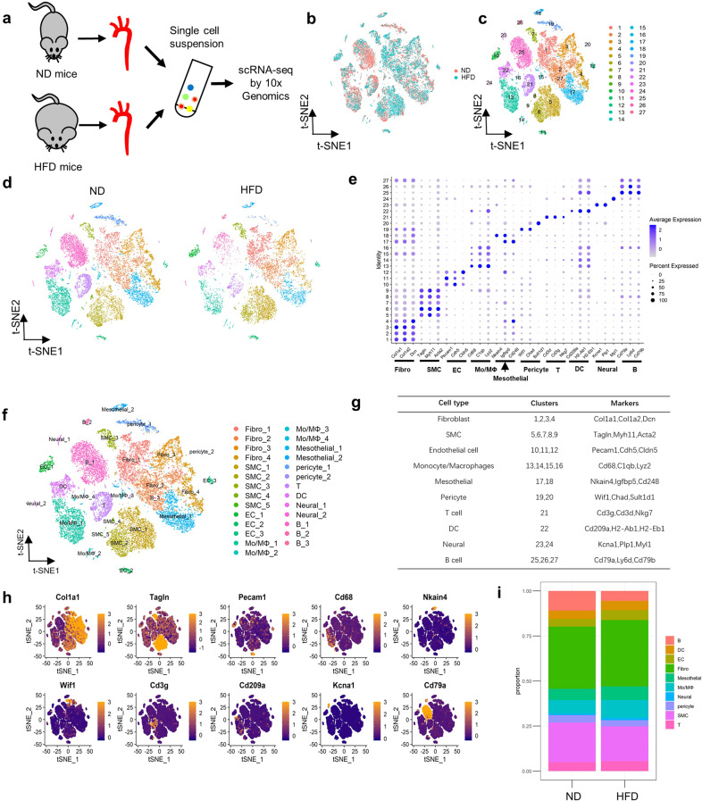
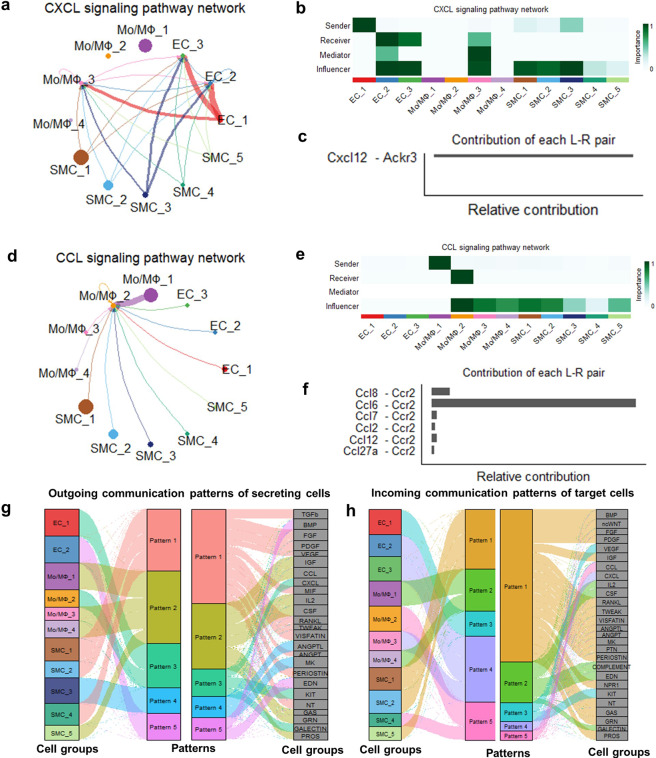
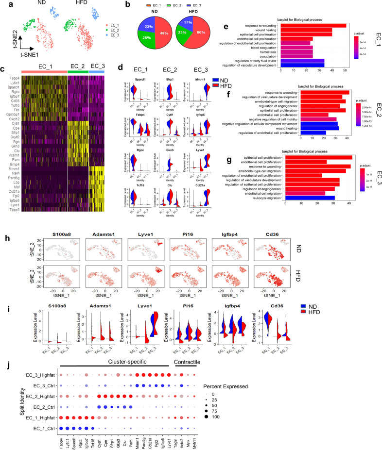
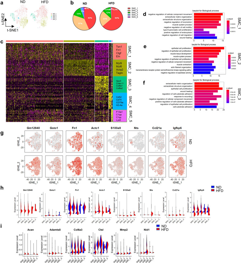
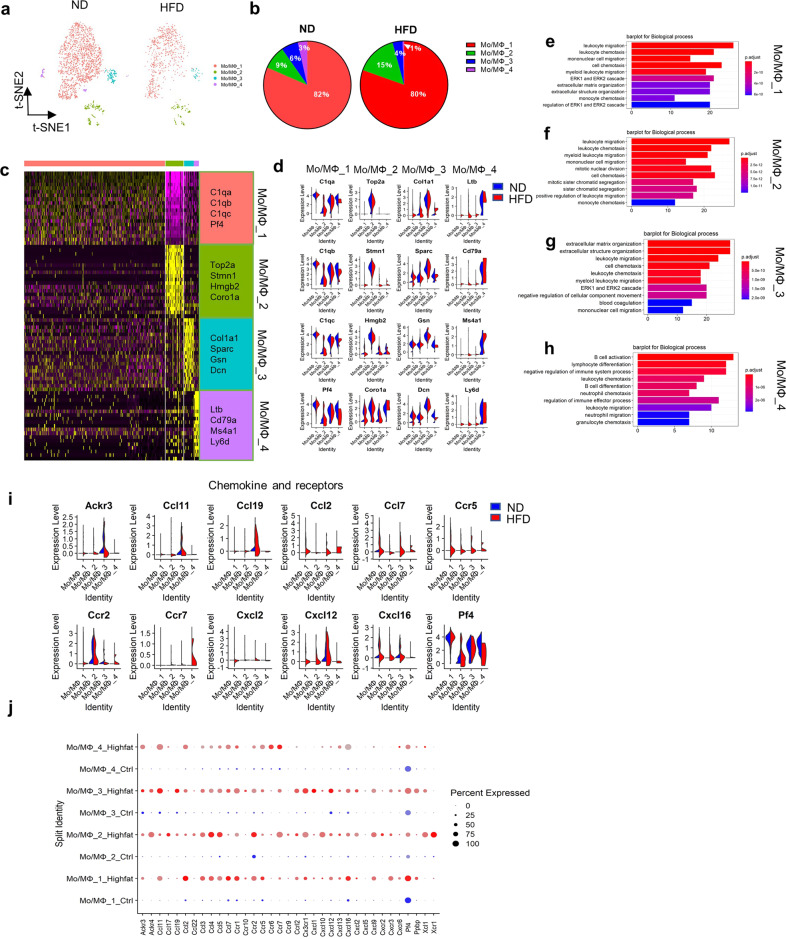
The aorta contains numerous cell types that contribute to vascular inflammation and thus the progression of aortic diseases. However, the heterogeneity and cellular composition of the ascending aorta in the setting of a high-fat diet (HFD) have not been fully assessed. We performed single-cell RNA sequencing on ascending aortas from mice fed a normal diet and mice fed a HFD. Unsupervised cluster analysis of the transcriptional profiles from 24,001 aortic cells identified 27 clusters representing 10 cell types: endothelial cells (ECs), fibroblasts, vascular smooth muscle cells (SMCs), immune cells (B cells, T cells, macrophages, and dendritic cells), mesothelial cells, pericytes, and neural cells. After HFD intake, subpopulations of endothelial cells with lipid transport and angiogenesis capacity and extensive expression of contractile genes were defined. In the HFD group, three major SMC subpopulations showed increased expression of extracellular matrix-degradation genes, and a synthetic SMC subcluster was proportionally increased. This increase was accompanied by upregulation of proinflammatory genes. Under HFD conditions, aortic-resident macrophage numbers were increased, and blood-derived macrophages showed the strongest expression of proinflammatory cytokines. Our study elucidates the nature and range of the cellular composition of the ascending aorta and increases understanding of the development and progression of aortic inflammatory disease.
主动脉包含许多有助于血管炎症的细胞类型,从而促进主动脉疾病的进展。然而,高脂肪饮食(HFD)条件下升主动脉的异质性和细胞组成尚未得到充分评估。我们对正常饮食喂养的小鼠和 HFD 喂养的小鼠的升主动脉进行了单细胞 RNA 测序。对 24001 个主动脉细胞的转录谱进行无监督聚类分析,鉴定出 27 个簇,代表 10 种细胞类型:内皮细胞(ECs)、成纤维细胞、血管平滑肌细胞(SMCs)、免疫细胞(B 细胞、T 细胞、巨噬细胞和树突状细胞)、间皮细胞、周细胞和神经细胞。在 HFD 摄入后,确定了具有脂质转运和血管生成能力以及广泛表达收缩基因的内皮细胞亚群。在 HFD 组中,三个主要的 SMC 亚群表现出细胞外基质降解基因的表达增加,并且合成 SMC 亚群成比例增加。这种增加伴随着促炎基因的上调。在 HFD 条件下,主动脉驻留巨噬细胞数量增加,并且血液衍生的巨噬细胞表现出最强的促炎细胞因子表达。我们的研究阐明了升主动脉的细胞组成的性质和范围,并增加了对主动脉炎症性疾病的发展和进展的理解。