Department of Radiology and Medical Imaging, University of Virginia, Charlottesville, VA 22901, USA.
Department of Medicine, University of Virginia, Charlottesville, VA 22901, USA.
Tomography. 2021 Sep 15;7(3):452-465. doi: 10.3390/tomography7030039.
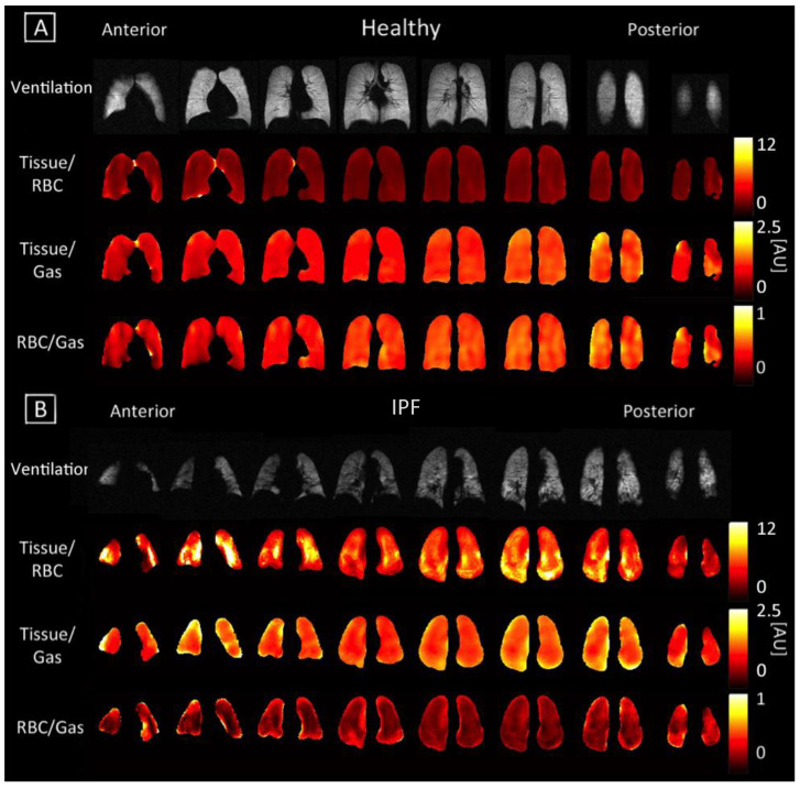
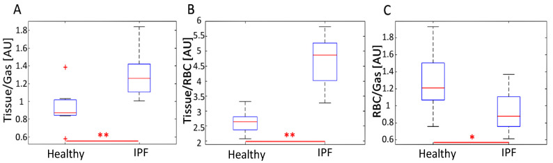
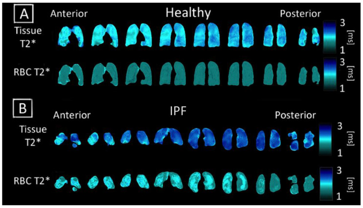
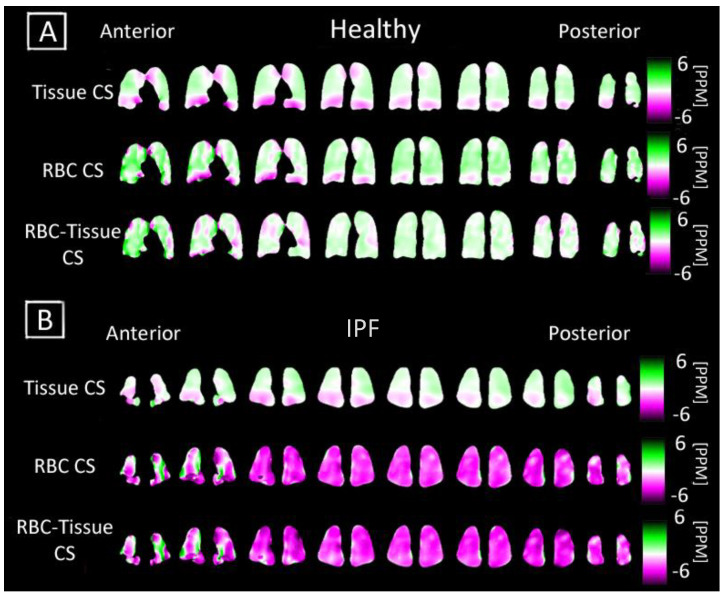
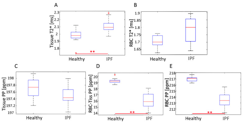
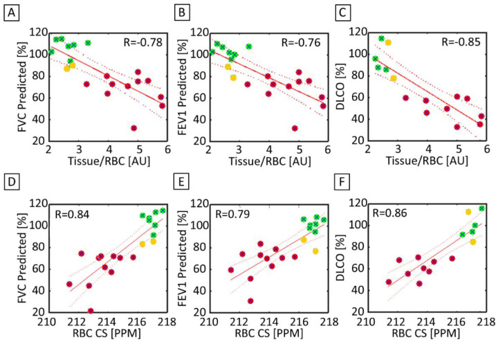
Idiopathic pulmonary fibrosis, a pattern of interstitial lung disease, is often clinically unpredictable in its progression. This paper presents hyperpolarized Xenon-129 chemical shift imaging as a noninvasive, nonradioactive method of probing lung physiology as well as anatomy to monitor subtle changes in subjects with IPF. Twenty subjects, nine healthy and eleven IPF, underwent HP Xe-129 ventilation MRI and 3D-SBCSI. Spirometry was performed on all subjects before imaging, and DLCO and hematocrit were measured in IPF subjects after imaging. Images were post-processed in MATLAB and segmented using ANTs. IPF subjects exhibited, on average, higher Tissue/Gas ratios and lower RBC/Gas ratios compared with healthy subjects, and quantitative maps were more heterogeneous in IPF subjects. The higher ratios are likely due to fibrosis and thickening of the pulmonary interstitium. T2* relaxation was longer in IPF subjects and corresponded with hematocrit scores, although the mechanism is not well understood. A lower chemical shift in the red blood cell spectroscopic peak correlated well with a higher Tissue/RBC ratio and may be explained by reduced blood oxygenation. Tissue/RBC also correlated well, spatially, with areas of fibrosis in HRCT images. These results may help us understand the underlying mechanism behind gas exchange impairment and disease progression.
特发性肺纤维化(一种间质性肺疾病模式)的进展在临床上常常难以预测。本文提出了氙气-129 化学位移成像作为一种非侵入性、非放射性的方法,用于探测肺生理和解剖结构,以监测特发性肺纤维化患者的细微变化。20 名受试者,9 名健康人和 11 名特发性肺纤维化患者,接受了 HP Xe-129 通气 MRI 和 3D-SBCSI 检查。所有受试者在成像前均进行了肺量计检查,成像后测量了特发性肺纤维化患者的 DLCO 和红细胞压积。图像在 MATLAB 中进行后处理,并使用 ANTs 进行分割。与健康受试者相比,特发性肺纤维化患者的组织/气体比平均更高,红细胞/气体比更低,并且特发性肺纤维化患者的定量图更不均匀。更高的比值可能是由于肺间质纤维化和增厚所致。特发性肺纤维化患者的 T2*弛豫时间较长,与红细胞压积分数相对应,尽管其机制尚不清楚。红细胞光谱峰的化学位移降低与组织/红细胞比值升高密切相关,这可能是由于血氧饱和度降低所致。组织/红细胞也与 HRCT 图像中纤维化区域在空间上很好地相关。这些结果可能有助于我们了解气体交换受损和疾病进展背后的潜在机制。