Wang Zeyu, Wang Haichen, Becker Ryan, Rufo Joseph, Yang Shujie, Mace Brian E, Wu Mengxi, Zou Jun, Laskowitz Daniel T, Huang Tony Jun
Department of Mechanical Engineering and Materials Science, Duke University, Durham, NC 27708 USA.
Department of Neurology, Duke University, Durham, NC 27708 USA.
Microsyst Nanoeng. 2021 Mar 3;7:20. doi: 10.1038/s41378-021-00244-3. eCollection 2021.
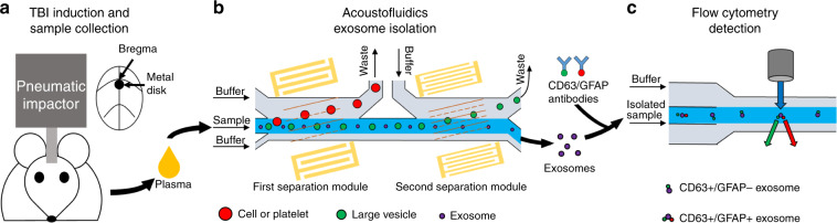
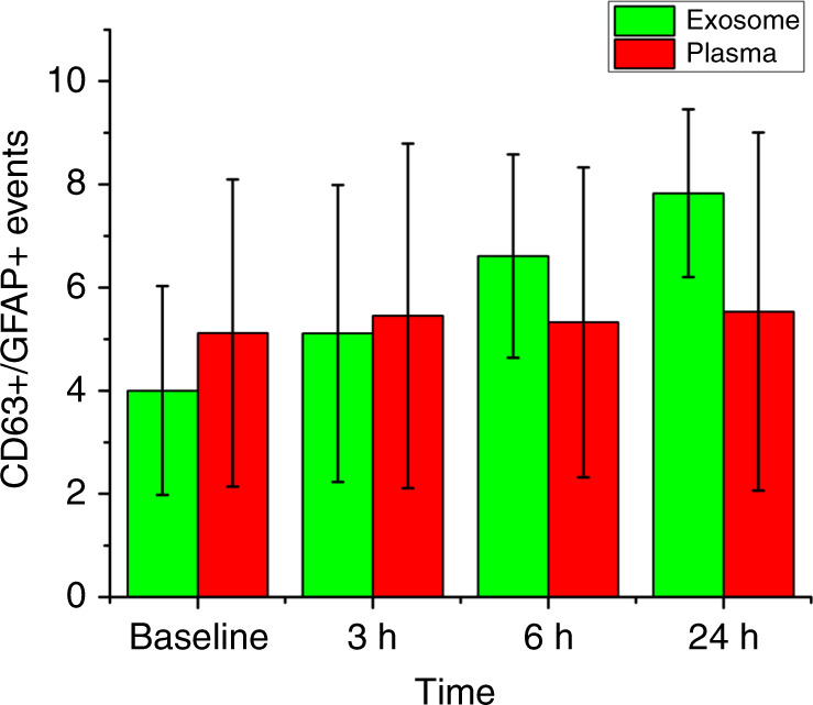
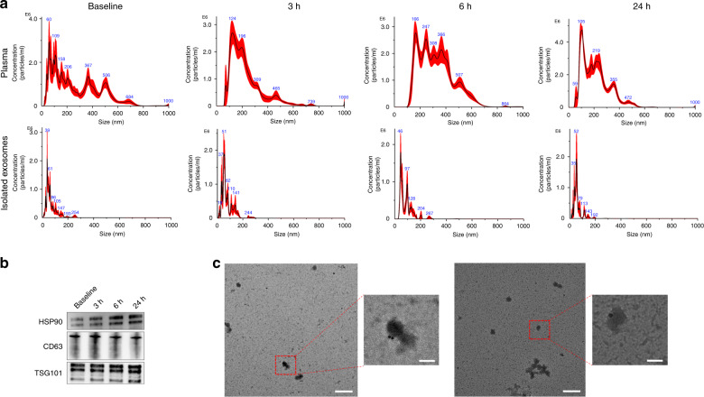
Traumatic brain injury (TBI) is a global cause of morbidity and mortality. Initial management and risk stratification of patients with TBI is made difficult by the relative insensitivity of screening radiographic studies as well as by the absence of a widely available, noninvasive diagnostic biomarker. In particular, a blood-based biomarker assay could provide a quick and minimally invasive process to stratify risk and guide early management strategies in patients with mild TBI (mTBI). Analysis of circulating exosomes allows the potential for rapid and specific identification of tissue injury. By applying acoustofluidic exosome separation-which uses a combination of microfluidics and acoustics to separate bioparticles based on differences in size and acoustic properties-we successfully isolated exosomes from plasma samples obtained from mice after TBI. Acoustofluidic isolation eliminated interference from other blood components, making it possible to detect exosomal biomarkers for TBI via flow cytometry. Flow cytometry analysis indicated that exosomal biomarkers for TBI increase in the first 24 h following head trauma, indicating the potential of using circulating exosomes for the rapid diagnosis of TBI. Elevated levels of TBI biomarkers were only detected in the samples separated via acoustofluidics; no changes were observed in the analysis of the raw plasma sample. This finding demonstrated the necessity of sample purification prior to exosomal biomarker analysis. Since acoustofluidic exosome separation can easily be integrated with downstream analysis methods, it shows great potential for improving early diagnosis and treatment decisions associated with TBI.
创伤性脑损伤(TBI)是全球范围内发病和死亡的原因。TBI患者的初始管理和风险分层因筛查影像学研究的相对不敏感性以及缺乏广泛可用的非侵入性诊断生物标志物而变得困难。特别是,基于血液的生物标志物检测可以提供一种快速且微创的方法来对轻度TBI(mTBI)患者进行风险分层并指导早期管理策略。循环外泌体的分析为快速、特异性地识别组织损伤提供了可能。通过应用声流体外泌体分离技术(该技术利用微流体和声学的组合,根据大小和声学特性的差异分离生物颗粒),我们成功地从小鼠TBI后的血浆样本中分离出了外泌体。声流体分离消除了其他血液成分的干扰,使得通过流式细胞术检测TBI的外泌体生物标志物成为可能。流式细胞术分析表明,TBI的外泌体生物标志物在头部创伤后的最初24小时内增加,这表明使用循环外泌体进行TBI快速诊断的潜力。仅在通过声流体分离的样本中检测到TBI生物标志物水平升高;在原始血浆样本的分析中未观察到变化。这一发现证明了在外泌体生物标志物分析之前进行样本纯化的必要性。由于声流体外泌体分离可以很容易地与下游分析方法整合,它在改善与TBI相关的早期诊断和治疗决策方面显示出巨大潜力。