Liao Chih-Kai, Fang Kuan-Min, Huang Hui-Ting, Chang Wen-Ruei, Chuang Chao-Chi, Tzeng Shun-Fen
Institute of Life Sciences, College of Bioscience and Biotechnology, National Cheng Kung University, Tainan 70101, Taiwan.
Department of Anatomy, Faculty of Medicine, Chung Shang Medical University, Taichung 40241, Taiwan.
Life (Basel). 2021 Sep 14;11(9):961. doi: 10.3390/life11090961.
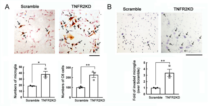
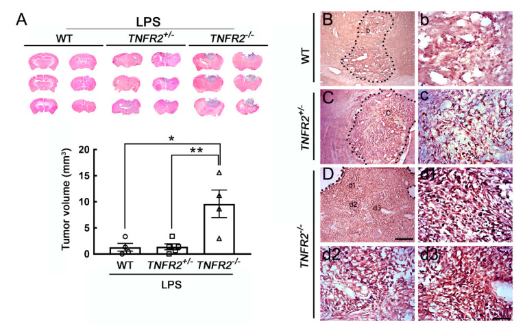
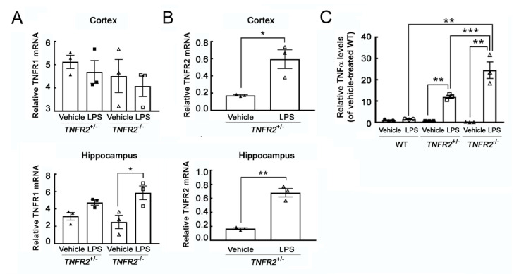
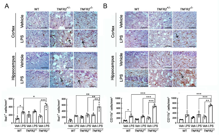
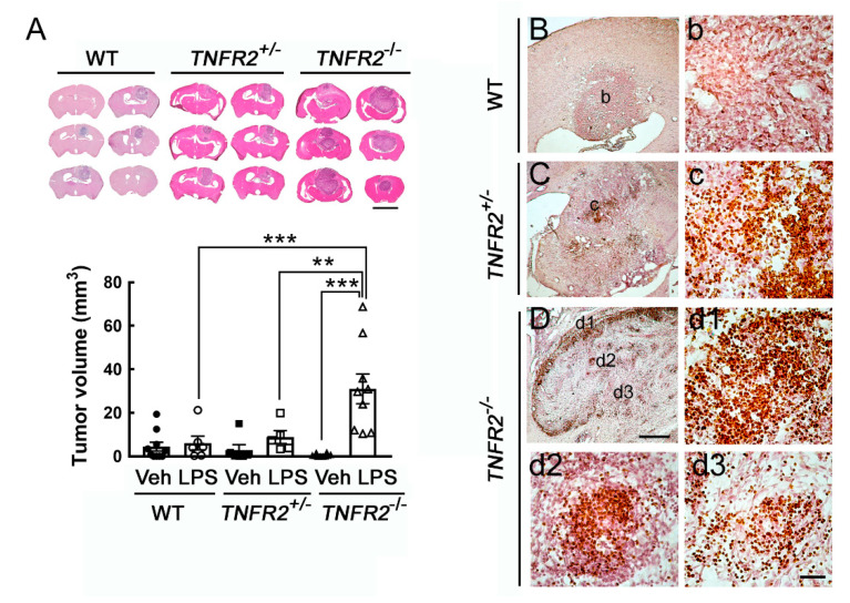
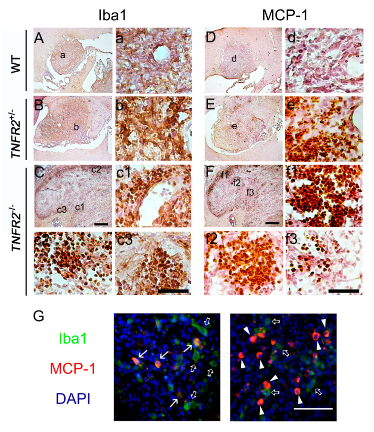
Despite the fact that accumulation of microglia, the resident macrophages of the central nervous system (CNS) are the main feature of glioblastoma, the role of microglia in the progression of glioma is still arguable. Based on the correlation of inflammation with tumor progression, in this study, we attempt to determine if peripheral inflammation aggravates glioma expansion and the activation of microglia associated with the tumor. Experimental animals were administered intraperitoneally by inflammagen lipopolysaccharide (LPS) for 7 days (LPS priming) before intracerebral implantation of glioma cells. Moreover, a reduced level of tumor necrosis factor receptor type 2 (TNFR2) that is restricted to immune cells, neurons, and microglia has been found in patients with glioblastoma through the clinic analysis of monocyte receptor expression. Thus, in addition to wildtype (WT) mice, heterogeneous TNFR2 gene deficiency () mice and homogeneous TNFR2 gene knockout () mice were used in this study. The results show that peripheral challenge by LPS, Iba1- or CD11b-microglia increase in numbers in the cortex and hippocampus of mice, when compared to WT or mice. We further conducted the intracerebral implantation of rodent glioma cells into the animals and found that the volumes of tumors formed by rat C6 glioma cells or mouse GL261 glioma cells were significantly larger in the cortex of mice when compared to that measured in LPS-primed WT or LPS-primed mice. Ki67-cells were exclusively clustered in the tumor of LPS-primed mice. Microglia were also extensively accumulated in the tumor formed in LPS-primed mice. Accordingly, our findings demonstrate that aggravation of microglia activation by peripheral inflammatory challenge and a loss of TNFR2 function might lead to the promotion of glioma growth.
尽管小胶质细胞(中枢神经系统(CNS)的常驻巨噬细胞)的积累是胶质母细胞瘤的主要特征,但小胶质细胞在胶质瘤进展中的作用仍存在争议。基于炎症与肿瘤进展的相关性,在本研究中,我们试图确定外周炎症是否会加剧胶质瘤的扩展以及与肿瘤相关的小胶质细胞的激活。在脑内植入胶质瘤细胞之前,对实验动物腹腔注射炎性介质脂多糖(LPS)7天(LPS预处理)。此外,通过对单核细胞受体表达的临床分析发现,胶质母细胞瘤患者中仅限于免疫细胞、神经元和小胶质细胞的肿瘤坏死因子受体2(TNFR2)水平降低。因此,除了野生型(WT)小鼠外,本研究还使用了异质性TNFR2基因缺陷()小鼠和同质性TNFR2基因敲除()小鼠。结果表明,与WT或小鼠相比,LPS对外周的刺激使小鼠皮质和海马中Iba1或CD11b小胶质细胞数量增加。我们进一步将啮齿类胶质瘤细胞脑内植入动物体内,发现与LPS预处理的WT或LPS预处理的小鼠相比,大鼠C6胶质瘤细胞或小鼠GL261胶质瘤细胞在小鼠皮质中形成的肿瘤体积显著更大。Ki67细胞仅聚集在LPS预处理的小鼠肿瘤中。小胶质细胞也广泛聚集在LPS预处理的小鼠形成的肿瘤中。因此,我们的研究结果表明,外周炎症刺激加剧小胶质细胞激活以及TNFR2功能丧失可能导致胶质瘤生长的促进。