Douglas Research Center, McGill University, Montréal, Canada.
University of California, San Francisco, USA.
Mol Brain. 2021 Sep 28;14(1):151. doi: 10.1186/s13041-021-00862-y.
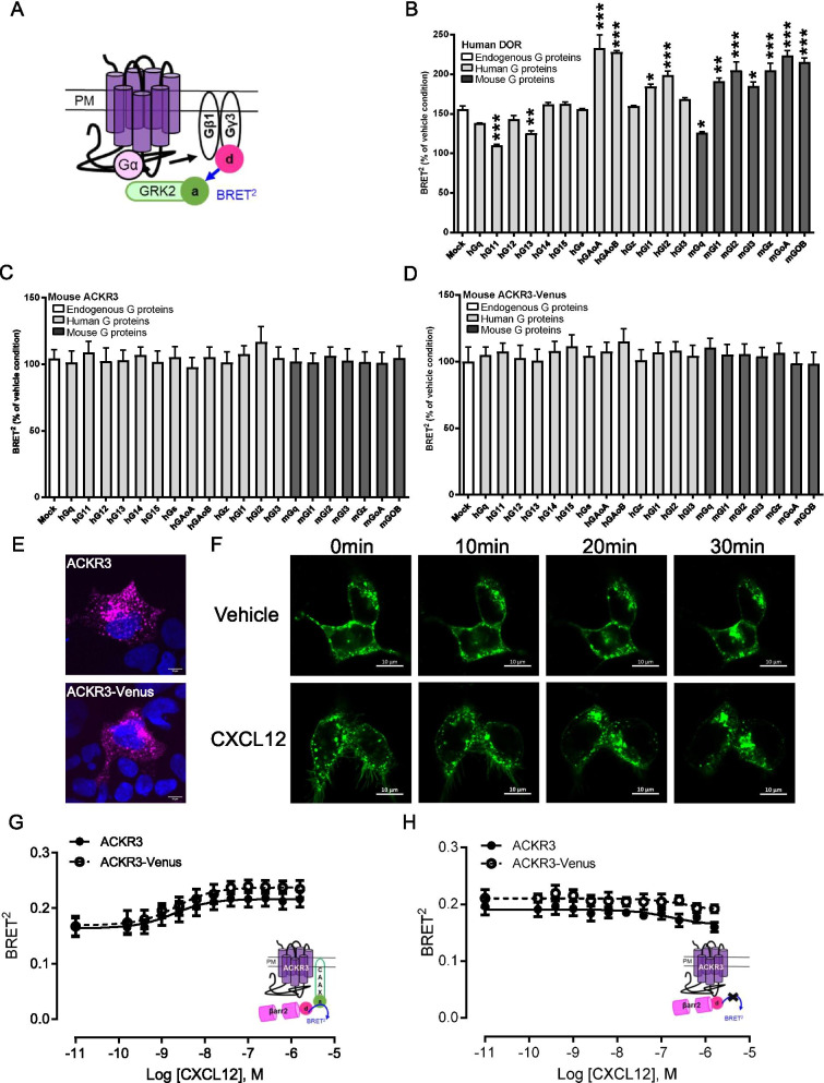
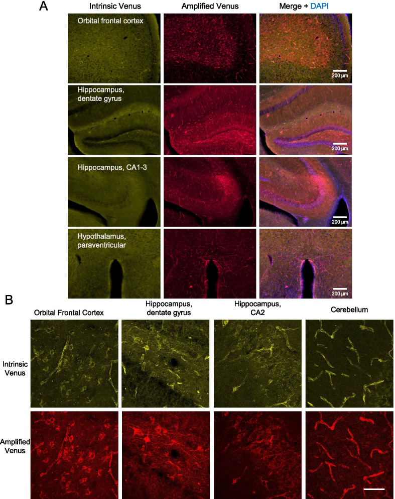
The atypical chemokine receptor 3, ACKR3, is a G protein-coupled receptor, which does not couple to G proteins but recruits βarrestins. At present, ACKR3 is considered a target for cancer and cardiovascular disorders, but less is known about the potential of ACKR3 as a target for brain disease. Further, mouse lines have been created to identify cells expressing the receptor, but there is no tool to visualize and study the receptor itself under physiological conditions. Here, we engineered a knock-in (KI) mouse expressing a functional ACKR3-Venus fusion protein to directly detect the receptor, particularly in the adult brain. In HEK-293 cells, native and fused receptors showed similar membrane expression, ligand induced trafficking and signaling profiles, indicating that the Venus fusion does not alter receptor signaling. We also found that ACKR3-Venus enables direct real-time monitoring of receptor trafficking using resonance energy transfer. In ACKR3-Venus knock-in mice, we found normal ACKR3 mRNA levels in the brain, suggesting intact gene transcription. We fully mapped receptor expression across 14 peripheral organs and 112 brain areas and found that ACKR3 is primarily localized to the vasculature in these tissues. In the periphery, receptor distribution aligns with previous reports. In the brain there is notable ACKR3 expression in endothelial vascular cells, hippocampal GABAergic interneurons and neuroblast neighboring cells. In conclusion, we have generated Ackr3-Venus knock-in mice with a traceable ACKR3 receptor, which will be a useful tool to the research community for interrogations about ACKR3 biology and related diseases.
非典型趋化因子受体 3(ACKR3)是一种 G 蛋白偶联受体,它不与 G 蛋白偶联,而是募集β-arrestin。目前,ACKR3 被认为是癌症和心血管疾病的靶点,但对于 ACKR3 作为脑部疾病靶点的潜力知之甚少。此外,已经创建了小鼠品系来鉴定表达该受体的细胞,但没有工具可在生理条件下可视化和研究受体本身。在这里,我们构建了一种表达功能性 ACKR3-Venus 融合蛋白的敲入(KI)小鼠,以直接检测受体,特别是在成年大脑中。在 HEK-293 细胞中,天然和融合受体显示出相似的膜表达、配体诱导的运输和信号转导特征,表明 Venus 融合不会改变受体信号转导。我们还发现 ACKR3-Venus 能够使用共振能量转移直接实时监测受体运输。在 ACKR3-Venus 敲入小鼠中,我们发现大脑中 ACKR3 的 mRNA 水平正常,表明基因转录完整。我们全面绘制了 14 个外周器官和 112 个脑区的受体表达图谱,发现 ACKR3 主要定位于这些组织的脉管系统。在外周组织中,受体分布与之前的报道一致。在大脑中,ACKR3 在血管内皮细胞、海马 GABA 能中间神经元和邻近神经母细胞中有明显表达。总之,我们已经生成了具有可追踪 ACKR3 受体的 Ackr3-Venus 敲入小鼠,这将是研究 ACKR3 生物学和相关疾病的有用工具。