Dipartimento di Medicina Veterinaria, Università di Sassari, Sassari, Italy.
Department of Zoology, University of Sargodha, Sargodha, Pakistan.
Parasit Vectors. 2021 Sep 28;14(1):505. doi: 10.1186/s13071-021-05013-9.
Gastrointestinal nematodes (GIN) are ubiquitous in small ruminant farming, representing a major health and production concern. Given their differences in pathogenicity and the current problems regarding anthelmintic resistance, specific diagnosis of GIN is of significant importance. At present, the most widely applied method for this entails culture and microscopic analysis of third-stage larvae, allowing for identification at least to the genus level. Overall, a variety of keys for microscopic analysis have been published, showing substantial variation. Given this fact, this study aimed to produce a practical and updated guide for the identification of infective ovine GIN larvae.
Using existing keys and protocols, a total of 173larvae of the most common species/genera of ovine GIN from pooled faecal samples from Sardinia (Italy) were identified and extracted, and further individual molecular identification was performed. Morphometric and morphological data as well as high-quality photographs were collected and combined to produce the final guide.
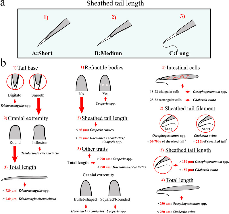
GIN microscopically and molecularly identified during this research include Trichostrongylus spp., Teladorsagia circumcincta, Haemonchus contortus, Cooperia curticei, and Chabertia ovina. Based on microscopic analysis, 73.5% of the larvae were correctly identified. Based on sheathed tail length, 91.8% were correctly classified into their respective preliminary groups.
It is crucial for the microscopic identification of infectious GIN larvae to examine each larva in its entirety and thus to take multiple characteristics into account to obtain an accurate diagnosis. However, a preliminary classification based on sheathed tail length (resulting in three groups: A, short; B, medium; C, long) was found to be effective. Further identification within group A can be achieved based on the presence of a cranial inflexion, caudal tubercles and full body measurements (Trichostrongylus spp. < 720 µm, T. circumcincta ≥ 720 µm). Larvae within group B can be differentiated based on sheathed tail morphometry (H. contortus > 65 µm, C. curticei ≤ 65 µm), the presence of cranial refractile bodies, total body length measurements (H. contortus ≤ 790 µm, C. curticei > 790 µm) and shape of the cranial extremity. Finally, all characteristics proposed for the differentiation between Oesophagostomum spp. and C. ovina larvae (group C) were found to have considerable restrictions.
胃肠道线虫(GIN)在小反刍动物养殖中普遍存在,是主要的健康和生产问题。鉴于它们在致病性方面的差异,以及目前驱虫剂耐药性方面的问题,对 GIN 进行特定诊断具有重要意义。目前,最广泛应用的方法是培养和第三期幼虫的显微镜分析,至少可以鉴定到属的水平。总的来说,已经发表了多种用于显微镜分析的关键方法,显示出很大的差异。鉴于这一事实,本研究旨在为鉴定感染性绵羊 GIN 幼虫提供实用和最新的指南。
使用现有的关键方法和协议,从撒丁岛(意大利)的混合粪便样本中鉴定和提取了总共 173 条最常见的绵羊 GIN 幼虫,并进行了进一步的个体分子鉴定。收集并结合了形态计量和形态学数据以及高质量的照片,以制作最终指南。
本研究中通过显微镜和分子鉴定的 GIN 包括 Trichostrongylus spp.、Teladorsagia circumcincta、Haemonchus contortus、Cooperia curticei 和 Chabertia ovina。根据显微镜分析,73.5%的幼虫得到了正确的鉴定。根据鞘尾长度,91.8%的幼虫被正确地分类到各自的初步组中。
对于传染性 GIN 幼虫的显微镜鉴定,必须检查每个幼虫的整体,从而考虑多个特征以获得准确的诊断。然而,发现基于鞘尾长度的初步分类(分为三组:A,短;B,中;C,长)是有效的。在组 A 中,可以进一步根据颅弯曲、尾端结节和全长测量(Trichostrongylus spp. < 720 µm,T. circumcincta ≥ 720 µm)进行鉴定。组 B 中的幼虫可以根据鞘尾形态计量学(H. contortus > 65 µm,C. curticei ≤ 65 µm)、颅反射体的存在、全长测量(H. contortus ≤ 790 µm,C. curticei > 790 µm)和颅端形状进行区分。最后,发现为区分 Oesophagostomum spp. 和 C. ovina 幼虫(组 C)而提出的所有特征都有很大的限制。