Taeger Johannes, Müller-Graff Franz Tassilo, Ilgen Lukas, Schendzielorz Phillip, Hagen Rudolf, Neun Tilman, Rak Kristen
Department of Oto-rhino-laryngology, Plastic, Aesthetic and Reconstructive Head and Neck Surgery and the Comprehensive Hearing Center, University of Wuerzburg, Wuerzburg, Germany.
Department of Diagnostic and Interventional Neuroradiology, University of Wuerzburg, Wuerzburg, Germany.
OTO Open. 2021 Sep 24;5(3):2473974X211045312. doi: 10.1177/2473974X211045312. eCollection 2021 Jul-Sep.
Growing interest in measuring the cochlear duct length (CDL) has emerged, since it can influence the selection of cochlear implant electrodes. Currently the measurements are performed with ionized radiation imaging. Only a few studies have explored CDL measurements in magnetic resonance imaging (MRI). Therefore, the presented study aims to fill this gap by estimating CDL in MRI and comparing it with multislice computed tomography (CT).
Retrospective data analyses of 42 cochleae.
Tertiary care medical center.
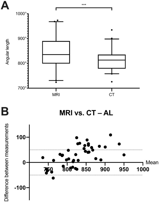
Diameter (A value) and width (B value) of the cochlea were measured in HOROS software. The CDL and the 2-turn length were determined by the elliptic circular approximation (ECA). In addition, the CDL, the 2-turn length, and the angular length were determined via HOROS software by the multiplanar reconstruction (MPR) method.
CDL values were significantly shorter in MRI by MPR ( = 1.38 mm, < .001) but not by ECA. Similar 2-turn length measurements were significantly lower in MRI by MPR ( = 1.67 mm) and ECA ( = 1.19 mm, both < .001). In contrast, angular length was significantly higher in MRI ( = 26.79°, < .001). When the values were set in relation to the frequencies of the cochlea, no clinically relevant differences were estimated (58 Hz at 28-mm CDL).
In the presented study, CDL was investigated in CT and MRI by using different approaches. Since no clinically relevant differences were found, diagnostics with radiation may be omitted prior to cochlear implantation; thus, a concept of radiation-free cochlear implantation could be established.
测量耳蜗管长度(CDL)的兴趣日益增加,因为它会影响人工耳蜗电极的选择。目前,测量是通过电离辐射成像进行的。只有少数研究探索了磁共振成像(MRI)中的CDL测量。因此,本研究旨在通过在MRI中估计CDL并将其与多层计算机断层扫描(CT)进行比较来填补这一空白。
对42个耳蜗进行回顾性数据分析。
三级医疗中心。
在HOROS软件中测量耳蜗的直径(A值)和宽度(B值)。通过椭圆-圆近似法(ECA)确定CDL和两圈长度。此外,通过HOROS软件,采用多平面重建(MPR)方法确定CDL、两圈长度和角长度。
通过MPR测量,MRI中的CDL值显著缩短( = 1.38 mm, <.001),但通过ECA测量则没有。通过MPR测量,MRI中类似的两圈长度测量值显著更低( = 1.67 mm),通过ECA测量也显著更低( = 1.19 mm,两者均 <.001)。相比之下,MRI中的角长度显著更高( = 26.79°, <.001)。当将这些值与耳蜗的频率相关联时,未估计出临床相关差异(在28 mm CDL时为58 Hz)。
在本研究中,采用不同方法在CT和MRI中对CDL进行了研究。由于未发现临床相关差异,人工耳蜗植入术前可省略放射诊断;因此,可以建立无辐射人工耳蜗植入的概念。