Seiger René, Hammerle Fabian P, Godbersen Godber M, Reed Murray B, Spurny-Dworak Benjamin, Handschuh Patricia, Klöbl Manfred, Unterholzner Jakob, Gryglewski Gregor, Vanicek Thomas, Lanzenberger Rupert
Department of Psychiatry and Psychotherapy, Medical University of Vienna, Vienna, Austria.
Front Neurosci. 2021 Sep 8;15:666000. doi: 10.3389/fnins.2021.666000. eCollection 2021.
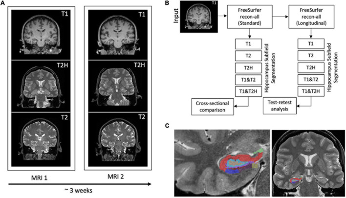
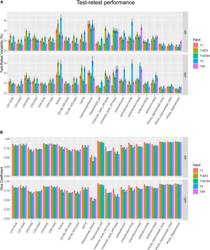
The accurate segmentation of magnetic resonance imaging (MRI) data is a crucial prerequisite for the reliable assessment of disease progression, patient stratification or the establishment of putative imaging biomarkers. This is especially important for the hippocampal formation, a brain area involved in memory formation and often affected by neurodegenerative or psychiatric diseases. FreeSurfer, a widely used automated segmentation software, offers hippocampal subfield delineation with multiple input options. While a single T1-weighted (T1) sequence is regularly used by most studies, it is also possible and advised to use a high-resolution T2-weighted (T2H) sequence or multispectral information. In this investigation it was determined whether there are differences in volume estimations depending on the input images and which combination of these deliver the most reliable results in each hippocampal subfield. 41 healthy participants (age = 25.2 years ± 4.2 SD) underwent two structural MRIs at three Tesla (time between scans: 23 days ± 11 SD) using three different structural MRI sequences, to test five different input configurations (T1, T2, T2H, T1 and T2, and T1 and T2H). We compared the different processing pipelines in a cross-sectional manner and assessed reliability using test-retest variability (%TRV) and the dice coefficient. Our analyses showed pronounced significant differences and large effect sizes between the processing pipelines in several subfields, such as the molecular layer (head), CA1 (head), hippocampal fissure, CA3 (head and body), fimbria and CA4 (head). The longitudinal analysis revealed that T1 and multispectral analysis (T1 and T2H) showed overall higher reliability across all subfields than T2H alone. However, the specific subfields had a substantial influence on the performance of segmentation results, regardless of the processing pipeline. Although T1 showed good test-retest metrics, results must be interpreted with caution, as a standard T1 sequence relies heavily on prior information of the atlas and does not take the actual fine structures of the hippocampus into account. For the most accurate segmentation, we advise the use of multispectral information by using a combination of T1 and high-resolution T2-weighted sequences or a T2 high-resolution sequence alone.
磁共振成像(MRI)数据的准确分割是可靠评估疾病进展、患者分层或建立假定影像生物标志物的关键前提。这对于海马结构尤为重要,海马是参与记忆形成的脑区,常受神经退行性疾病或精神疾病影响。FreeSurfer是一款广泛使用的自动分割软件,提供多种输入选项用于海马亚区描绘。虽然大多数研究通常使用单个T1加权(T1)序列,但使用高分辨率T2加权(T2H)序列或多光谱信息也是可行且被建议的。在本研究中,确定了根据输入图像进行体积估计时是否存在差异,以及这些图像的哪种组合在每个海马亚区能产生最可靠的结果。41名健康参与者(年龄 = 25.2岁±4.2标准差)在3特斯拉磁场下接受了两次结构MRI扫描(扫描间隔时间:23天±11标准差),使用三种不同的结构MRI序列,以测试五种不同的输入配置(T1、T2、T2H、T1和T2以及T1和T2H)。我们以横断面方式比较了不同的处理流程,并使用重测变异性(%TRV)和骰子系数评估可靠性。我们的分析表明,在几个亚区,如分子层(头部)、CA1(头部)、海马裂、CA3(头部和体部)、伞和CA4(头部),处理流程之间存在显著差异和较大效应量。纵向分析显示,T1和多光谱分析(T1和T2H)在所有亚区的总体可靠性高于单独的T2H。然而,无论处理流程如何,特定亚区对分割结果的性能有重大影响。虽然T1显示出良好的重测指标,但结果必须谨慎解读,因为标准T1序列严重依赖图谱的先验信息,未考虑海马的实际精细结构。为了实现最准确的分割,我们建议通过结合T1和高分辨率T2加权序列或单独使用T2高分辨率序列来使用多光谱信息。