• 文献检索
  • 文档翻译
  • 深度研究
  • 学术资讯
  • Suppr Zotero 插件Zotero 插件
  • 邀请有礼
  • 套餐&价格
  • 历史记录
应用&插件
Suppr Zotero 插件Zotero 插件浏览器插件Mac 客户端Windows 客户端微信小程序
定价
高级版会员购买积分包购买API积分包
服务
文献检索文档翻译深度研究API 文档MCP 服务
关于我们
关于 Suppr公司介绍联系我们用户协议隐私条款
关注我们

Suppr 超能文献

核心技术专利:CN118964589B侵权必究
粤ICP备2023148730 号-1Suppr @ 2026

文献检索

告别复杂PubMed语法,用中文像聊天一样搜索,搜遍4000万医学文献。AI智能推荐,让科研检索更轻松。

立即免费搜索

文件翻译

保留排版,准确专业,支持PDF/Word/PPT等文件格式,支持 12+语言互译。

免费翻译文档

深度研究

AI帮你快速写综述,25分钟生成高质量综述,智能提取关键信息,辅助科研写作。

立即免费体验

基于内镜超声图像的神经网络鉴别胃肠道间质瘤与平滑肌瘤。

Differentiating Gastrointestinal Stromal Tumors from Leiomyomas Using a Neural Network Trained on Endoscopic Ultrasonography Images.

机构信息

Division of Gastroenterology, Bezmialem Vakif University, Istanbul, Turkey.

Management Information Systems Department, School of Business and Management Science, Istanbul Medipol University, Istanbul, Turkey.

出版信息

Dig Dis. 2022;40(4):427-435. doi: 10.1159/000520032. Epub 2021 Oct 7.

DOI:10.1159/000520032
PMID:34619683
原文链接:https://pmc.ncbi.nlm.nih.gov/articles/PMC9393815/
Abstract

BACKGROUND

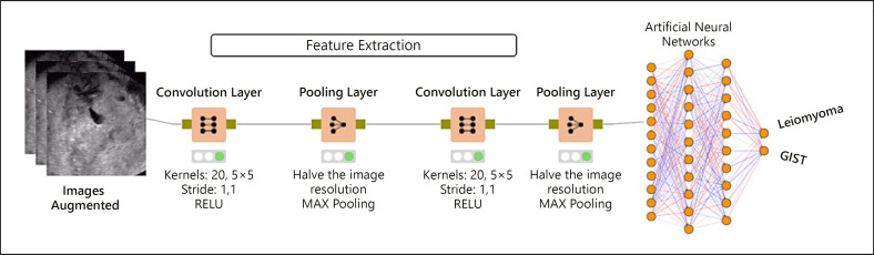
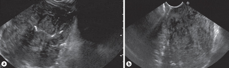
Endoscopic ultrasonography (EUS) is crucial to diagnose and evaluate gastrointestinal mesenchymal tumors (GIMTs). However, EUS-guided biopsy does not always differentiate gastrointestinal stromal tumors (GISTs) from leiomyomas. We evaluated the ability of a convolutional neural network (CNN) to differentiate GISTs from leiomyomas using EUS images. The conventional EUS features of GISTs were also compared with leiomyomas.

PATIENTS AND METHODS

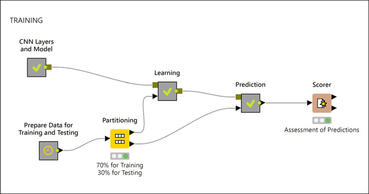
Patients who underwent EUS for evaluation of upper GIMTs between 2010 and 2020 were retrospectively reviewed, and 145 patients (73 women and 72 men; mean age 54.8 ± 13.5 years) with GISTs (n = 109) or leiomyomas (n = 36), confirmed by immunohistochemistry, were included. A total of 978 images collected from 100 patients were used to train and test the CNN system, and 384 images from 45 patients were used for validation. EUS images were also evaluated by an EUS expert for comparison with the CNN system.

RESULTS

The sensitivity, specificity, and accuracy of the CNN system for diagnosis of GIST were 92.0%, 64.3%, and 86.98% for the validation dataset, respectively. In contrast, the sensitivity, specificity, and accuracy of the EUS expert interpretations were 60.5%, 74.3%, and 63.0%, respectively. Concerning EUS features, only higher echogenicity was an independent and significant factor for differentiating GISTs from leiomyomas (p < 0.05).

CONCLUSIONS

The CNN system could diagnose GIMTs with higher accuracy than an EUS expert and could be helpful in differentiating GISTs from leiomyomas. A higher echogenicity may also aid in differentiation.

摘要

背景

内镜超声检查(EUS)对于诊断和评估胃肠道间质肿瘤(GIMTs)至关重要。然而,EUS 引导下的活检并不总能区分胃肠道间质瘤(GISTs)和平滑肌瘤。我们评估了卷积神经网络(CNN)使用 EUS 图像区分 GISTs 和平滑肌瘤的能力。还比较了 GISTs 的传统 EUS 特征与平滑肌瘤。

患者和方法

回顾性分析 2010 年至 2020 年间因上 GIMTs 而行 EUS 评估的患者,纳入 145 例(73 名女性和 72 名男性;平均年龄 54.8 ± 13.5 岁),经免疫组织化学证实为 GIST(n = 109)或平滑肌瘤(n = 36)。从 100 名患者中采集了 978 张图像用于训练和测试 CNN 系统,从 45 名患者中采集了 384 张图像用于验证。EUS 图像也由 EUS 专家进行评估,以便与 CNN 系统进行比较。

结果

CNN 系统对验证数据集 GIST 的诊断的敏感性、特异性和准确性分别为 92.0%、64.3%和 86.98%。相比之下,EUS 专家解释的敏感性、特异性和准确性分别为 60.5%、74.3%和 63.0%。关于 EUS 特征,只有更高的回声强度是区分 GISTs 和平滑肌瘤的独立且重要的因素(p < 0.05)。

结论

CNN 系统可以比 EUS 专家更准确地诊断 GIMTs,并且有助于区分 GISTs 和平滑肌瘤。回声强度较高也可能有助于区分。

https://cdn.ncbi.nlm.nih.gov/pmc/blobs/663e/9393815/b0273a25e6d0/ddi-0040-0427-g04.jpg
https://cdn.ncbi.nlm.nih.gov/pmc/blobs/663e/9393815/5c48481e63dc/ddi-0040-0427-g01.jpg
https://cdn.ncbi.nlm.nih.gov/pmc/blobs/663e/9393815/57b91c7cc55d/ddi-0040-0427-g02.jpg
https://cdn.ncbi.nlm.nih.gov/pmc/blobs/663e/9393815/9f97765ba09d/ddi-0040-0427-g03.jpg
https://cdn.ncbi.nlm.nih.gov/pmc/blobs/663e/9393815/b0273a25e6d0/ddi-0040-0427-g04.jpg

相似文献

1
Differentiating Gastrointestinal Stromal Tumors from Leiomyomas Using a Neural Network Trained on Endoscopic Ultrasonography Images.基于内镜超声图像的神经网络鉴别胃肠道间质瘤与平滑肌瘤。
Dig Dis. 2022;40(4):427-435. doi: 10.1159/000520032. Epub 2021 Oct 7.
2
Differentiating Gastrointestinal Stromal Tumors From Leiomyomas of Upper Digestive Tract Using Convolutional Neural Network Model by Endoscopic Ultrasonography.利用卷积神经网络模型通过内镜超声对上消化道平滑肌肿瘤与胃肠道间质瘤进行鉴别。
J Clin Gastroenterol. 2024 Jul 1;58(6):574-579. doi: 10.1097/MCG.0000000000001907.
3
Convolutional neural network-based object detection model to identify gastrointestinal stromal tumors in endoscopic ultrasound images.基于卷积神经网络的目标检测模型用于在内镜超声图像中识别胃肠道间质瘤。
J Gastroenterol Hepatol. 2021 Dec;36(12):3387-3394. doi: 10.1111/jgh.15653. Epub 2021 Aug 16.
4
Clinicopathological characteristics of gastrointestinal mesenchymal tumors and diagnostic value of endoscopic ultrasonography.胃肠道间质瘤的临床病理特征及内镜超声检查的诊断价值
J Gastroenterol Hepatol. 2008 Aug;23(8 Pt 2):e318-24. doi: 10.1111/j.1440-1746.2008.05322.x. Epub 2008 May 26.
5
Deep learning analysis for differential diagnosis and risk classification of gastrointestinal tumors.深度学习分析在胃肠道肿瘤的鉴别诊断和风险分类中的应用。
Scand J Gastroenterol. 2024 Aug;59(8):925-932. doi: 10.1080/00365521.2024.2368241. Epub 2024 Jul 1.
6
Is it possible to differentiate gastric GISTs from gastric leiomyomas by EUS?是否可以通过超声内镜(EUS)区分胃间质瘤(GISTs)和胃平滑肌瘤?
World J Gastroenterol. 2009 Jul 21;15(27):3376-81. doi: 10.3748/wjg.15.3376.
7
Application of A Convolutional Neural Network in The Diagnosis of Gastric Mesenchymal Tumors on Endoscopic Ultrasonography Images.卷积神经网络在内镜超声图像胃间质瘤诊断中的应用
J Clin Med. 2020 Sep 29;9(10):3162. doi: 10.3390/jcm9103162.
8
Digital image analysis-based scoring system for endoscopic ultrasonography is useful in predicting gastrointestinal stromal tumors.基于数字图像分析的内镜超声评分系统有助于预测胃肠道间质瘤。
Gastric Cancer. 2019 Sep;22(5):980-987. doi: 10.1007/s10120-019-00928-w. Epub 2019 Feb 18.
9
A combined radiomic model distinguishing GISTs from leiomyomas and schwannomas in the stomach based on endoscopic ultrasonography images.基于内镜超声图像的联合放射组学模型鉴别胃间质瘤、平滑肌瘤和神经鞘瘤。
J Appl Clin Med Phys. 2023 Jul;24(7):e14023. doi: 10.1002/acm2.14023. Epub 2023 May 11.
10
Value of artificial intelligence with novel tumor tracking technology in the diagnosis of gastric submucosal tumors by contrast-enhanced harmonic endoscopic ultrasonography.新型肿瘤跟踪技术的人工智能在对比增强谐波内镜超声诊断胃黏膜下肿瘤中的价值。
J Gastroenterol Hepatol. 2022 May;37(5):841-846. doi: 10.1111/jgh.15780. Epub 2022 Jan 31.

引用本文的文献

1
Multimodal artificial intelligence for subepithelial lesion classification and characterization: a multicenter comparative study (with video).用于上皮下病变分类和特征描述的多模态人工智能:一项多中心比较研究(附视频)
BMC Med Inform Decis Mak. 2025 Aug 14;25(1):307. doi: 10.1186/s12911-025-03147-9.
2
The role of artificial intelligence and deep learning in determining the histopathological grade of pancreatic neuroendocrine tumors by using EUS images.人工智能和深度学习在利用超声内镜(EUS)图像确定胰腺神经内分泌肿瘤组织病理学分级中的作用。
Endosc Ultrasound. 2025 Mar-Apr;14(2):48-56. doi: 10.1097/eus.0000000000000113. Epub 2025 May 2.
3

本文引用的文献

1
Use of Artificial Intelligence in the Prediction of Malignant Potential of Gastric Gastrointestinal Stromal Tumors.人工智能在胃胃肠间质瘤恶性潜能预测中的应用。
Dig Dis Sci. 2022 Jan;67(1):273-281. doi: 10.1007/s10620-021-06830-9. Epub 2021 Feb 6.
2
Application of A Convolutional Neural Network in The Diagnosis of Gastric Mesenchymal Tumors on Endoscopic Ultrasonography Images.卷积神经网络在内镜超声图像胃间质瘤诊断中的应用
J Clin Med. 2020 Sep 29;9(10):3162. doi: 10.3390/jcm9103162.
3
Efficacy of endoscopic ultrasound with artificial intelligence for the diagnosis of gastrointestinal stromal tumors.
A Lightweight Machine Learning Model for High Precision Gastrointestinal Stromal Tumors Identification.
一种用于高精度胃肠道间质瘤识别的轻量级机器学习模型。
Bioengineering (Basel). 2025 Apr 3;12(4):381. doi: 10.3390/bioengineering12040381.
4
An artificial intelligence model utilizing endoscopic ultrasonography for differentiating small and micro gastric stromal tumors from gastric leiomyomas.一种利用内镜超声检查区分小的和微小的胃间质瘤与胃平滑肌瘤的人工智能模型。
BMC Gastroenterol. 2025 Apr 9;25(1):237. doi: 10.1186/s12876-025-03825-y.
5
Differential diagnosis of gastrointestinal stromal tumors versus leiomyomas by special stains.特殊染色对胃肠道间质瘤与平滑肌瘤的鉴别诊断。
BMC Gastroenterol. 2024 Nov 21;24(1):418. doi: 10.1186/s12876-024-03511-5.
6
Artificial Intelligence-Based Diagnosis of Gastric Mesenchymal Tumors Using Digital Endosonography Image Analysis.基于人工智能的胃间质瘤数字超声内镜图像分析诊断
J Clin Med. 2024 Jun 26;13(13):3725. doi: 10.3390/jcm13133725.
7
Convolutional neural network applied to preoperative venous-phase CT images predicts risk category in patients with gastric gastrointestinal stromal tumors.卷积神经网络应用于术前静脉期 CT 图像预测胃胃肠道间质瘤患者的风险类别。
BMC Cancer. 2024 Mar 1;24(1):280. doi: 10.1186/s12885-024-11962-y.
8
Endoscopic ultrasound artificial intelligence-assisted for prediction of gastrointestinal stromal tumors diagnosis: A systematic review and meta-analysis.内镜超声人工智能辅助预测胃肠道间质瘤诊断:一项系统评价和荟萃分析。
World J Gastrointest Endosc. 2023 Aug 16;15(8):528-539. doi: 10.4253/wjge.v15.i8.528.
9
Current status of artificial intelligence analysis for the treatment of pancreaticobiliary diseases using endoscopic ultrasonography and endoscopic retrograde cholangiopancreatography.使用内镜超声和内镜逆行胰胆管造影术进行人工智能分析治疗胰胆疾病的现状
DEN Open. 2023 Jun 30;4(1):e267. doi: 10.1002/deo2.267. eCollection 2024 Apr.
10
Artificial intelligence in endoscopic ultrasonography: risk stratification of gastric gastrointestinal stromal tumors.人工智能在内镜超声检查中的应用:胃胃肠道间质瘤的风险分层
Therap Adv Gastroenterol. 2023 May 30;16:17562848231177156. doi: 10.1177/17562848231177156. eCollection 2023.
人工智能辅助内镜超声对胃肠道间质瘤的诊断效能
J Gastroenterol. 2020 Dec;55(12):1119-1126. doi: 10.1007/s00535-020-01725-4. Epub 2020 Sep 11.
4
EUS-guided fine-needle biopsy sampling versus FNA in the diagnosis of subepithelial lesions: a large multicenter study.EUS 引导下的细针活检与 FNA 在诊断黏膜下病变中的对比:一项大型多中心研究。
Gastrointest Endosc. 2020 Jul;92(1):108-119.e3. doi: 10.1016/j.gie.2020.02.021. Epub 2020 Feb 25.
5
Superiority of mucosal incision-assisted biopsy over ultrasound-guided fine needle aspiration biopsy in diagnosing small gastric subepithelial lesions: a propensity score matching analysis.黏膜切口辅助活检在诊断小胃黏膜下病变中的优越性:一项倾向评分匹配分析。
BMC Gastroenterol. 2020 Jan 21;20(1):19. doi: 10.1186/s12876-020-1170-2.
6
The role of EUS and EUS-guided FNA in the management of subepithelial lesions of the esophagus: A large, single-center experience.超声内镜及超声内镜引导下细针穿刺在食管上皮下病变管理中的作用:一项大型单中心经验。
Endosc Ultrasound. 2017 Sep-Oct;6(5):308-316. doi: 10.4103/2303-9027.155772.
7
Accuracy of a scoring system for the differential diagnosis of common gastric subepithelial tumors based on endoscopic ultrasonography.基于内镜超声的常见胃黏膜下肿瘤鉴别诊断评分系统的准确性。
J Dig Dis. 2013 Dec;14(12):647-53. doi: 10.1111/1751-2980.12099.
8
Concordance of endoscopic ultrasonography-guided fine needle aspiration diagnosis with the final diagnosis in subepithelial lesions.内镜超声引导下细针穿刺诊断与上皮下病变最终诊断的一致性。
Clin Endosc. 2013 Jul;46(4):379-83. doi: 10.5946/ce.2013.46.4.379. Epub 2013 Jul 31.
9
Is it possible to differentiate gastric GISTs from gastric leiomyomas by EUS?是否可以通过超声内镜(EUS)区分胃间质瘤(GISTs)和胃平滑肌瘤?
World J Gastroenterol. 2009 Jul 21;15(27):3376-81. doi: 10.3748/wjg.15.3376.
10
Gastrointestinal stromal tumors: pathology and prognosis at different sites.胃肠道间质瘤:不同部位的病理学与预后
Semin Diagn Pathol. 2006 May;23(2):70-83. doi: 10.1053/j.semdp.2006.09.001.