Materials and Structural Analysis, Thermo Fisher Scientific, Achtseweg Noord 5, 5651 GG Eindhoven, Netherlands.
School of Biosciences, University of Sheffield, Sheffield S10 2TN, U.K.
Biochem J. 2021 Nov 12;478(21):3923-3937. doi: 10.1042/BCJ20210696.
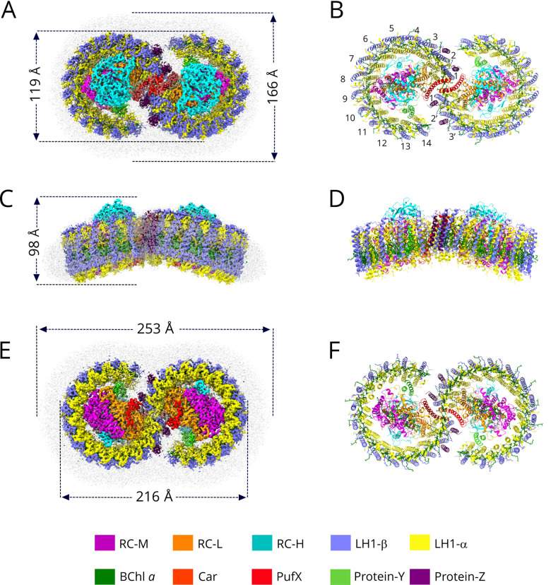
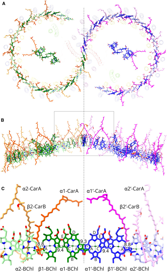
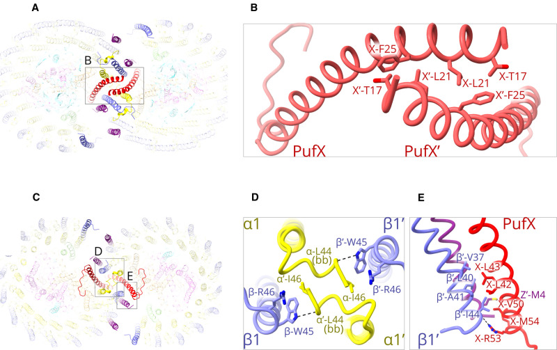
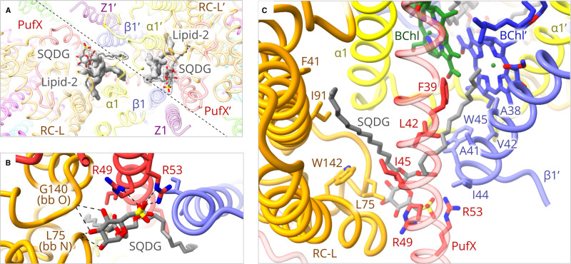
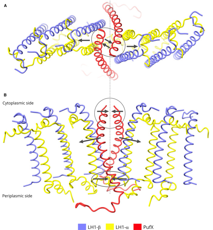
The dimeric reaction centre light-harvesting 1 (RC-LH1) core complex of Rhodobacter sphaeroides converts absorbed light energy to a charge separation, and then it reduces a quinone electron and proton acceptor to a quinol. The angle between the two monomers imposes a bent configuration on the dimer complex, which exerts a major influence on the curvature of the membrane vesicles, known as chromatophores, where the light-driven photosynthetic reactions take place. To investigate the dimerisation interface between two RC-LH1 monomers, we determined the cryogenic electron microscopy structure of the dimeric complex at 2.9 Å resolution. The structure shows that each monomer consists of a central RC partly enclosed by a 14-subunit LH1 ring held in an open state by PufX and protein-Y polypeptides, thus enabling quinones to enter and leave the complex. Two monomers are brought together through N-terminal interactions between PufX polypeptides on the cytoplasmic side of the complex, augmented by two novel transmembrane polypeptides, designated protein-Z, that bind to the outer faces of the two central LH1 β polypeptides. The precise fit at the dimer interface, enabled by PufX and protein-Z, by C-terminal interactions between opposing LH1 αβ subunits, and by a series of interactions with a bound sulfoquinovosyl diacylglycerol lipid, bring together each monomer creating an S-shaped array of 28 bacteriochlorophylls. The seamless join between the two sets of LH1 bacteriochlorophylls provides a path for excitation energy absorbed by one half of the complex to migrate across the dimer interface to the other half.
球形红杆菌的二聚反应中心光捕获 1(RC-LH1)核心复合物将吸收的光能转化为电荷分离,然后还原醌电子和质子受体为醌醇。两个单体之间的角度使二聚体复合物呈弯曲构象,这对称为类囊体的膜囊泡的曲率产生重大影响,光驱动的光合作用发生在类囊体中。为了研究两个 RC-LH1 单体之间的二聚化界面,我们确定了 2.9Å 分辨率的二聚复合物的低温电子显微镜结构。该结构表明,每个单体由一个被 14 个亚基 LH1 环部分包围的中央 RC 组成,LH1 环由 PufX 和蛋白-Y 多肽保持在开放状态,从而使醌能够进入和离开复合物。两个单体通过复合物细胞质侧的 PufX 多肽之间的 N 端相互作用聚集在一起,辅之以两个新的跨膜多肽,称为蛋白-Z,它们与两个中央 LH1 β多肽的外表面结合。通过 PufX 和蛋白-Z、相反的 LH1αβ 亚基之间的 C 端相互作用以及与结合的磺基奎诺二酰甘油脂质的一系列相互作用,在二聚体界面实现精确匹配,将每个单体聚集在一起,形成 28 个细菌叶绿素的 S 形排列。两组 LH1 细菌叶绿素之间的无缝连接为复合物一半吸收的激发能提供了一条穿过二聚体界面迁移到另一半的途径。