• 文献检索
  • 文档翻译
  • 深度研究
  • 学术资讯
  • Suppr Zotero 插件Zotero 插件
  • 邀请有礼
  • 套餐&价格
  • 历史记录
应用&插件
Suppr Zotero 插件Zotero 插件浏览器插件Mac 客户端Windows 客户端微信小程序
定价
高级版会员购买积分包购买API积分包
服务
文献检索文档翻译深度研究API 文档MCP 服务
关于我们
关于 Suppr公司介绍联系我们用户协议隐私条款
关注我们

Suppr 超能文献

核心技术专利:CN118964589B侵权必究
粤ICP备2023148730 号-1Suppr @ 2026

文献检索

告别复杂PubMed语法,用中文像聊天一样搜索,搜遍4000万医学文献。AI智能推荐,让科研检索更轻松。

立即免费搜索

文件翻译

保留排版,准确专业,支持PDF/Word/PPT等文件格式,支持 12+语言互译。

免费翻译文档

深度研究

AI帮你快速写综述,25分钟生成高质量综述,智能提取关键信息,辅助科研写作。

立即免费体验

无分割、自动化、血管矢量化。

Segmentation-Less, Automated, Vascular Vectorization.

机构信息

Department of Biomedical Engineering, The University of Texas, Austin, Texas, United States of America.

Institute for Neuroscience, The University of Texas, Austin, Texas, United States of America.

出版信息

PLoS Comput Biol. 2021 Oct 8;17(10):e1009451. doi: 10.1371/journal.pcbi.1009451. eCollection 2021 Oct.

DOI:10.1371/journal.pcbi.1009451
PMID:34624013
原文链接:https://pmc.ncbi.nlm.nih.gov/articles/PMC8528315/
Abstract

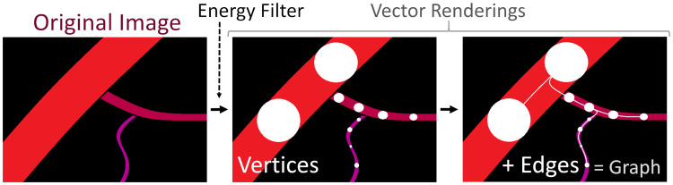
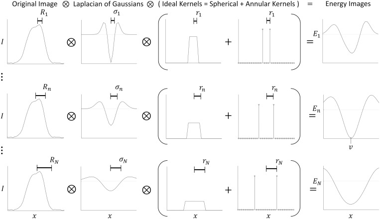
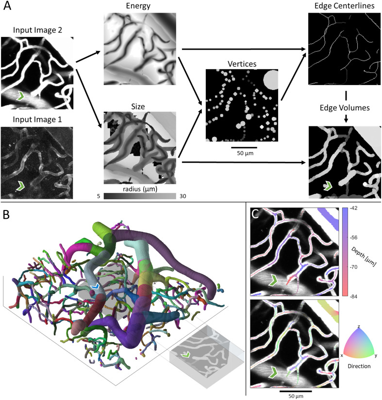
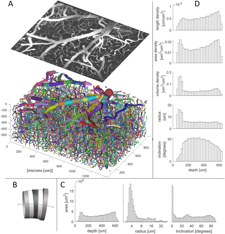
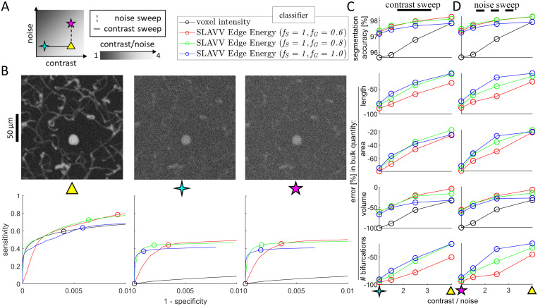
Recent advances in two-photon fluorescence microscopy (2PM) have allowed large scale imaging and analysis of blood vessel networks in living mice. However, extracting network graphs and vector representations for the dense capillary bed remains a bottleneck in many applications. Vascular vectorization is algorithmically difficult because blood vessels have many shapes and sizes, the samples are often unevenly illuminated, and large image volumes are required to achieve good statistical power. State-of-the-art, three-dimensional, vascular vectorization approaches often require a segmented (binary) image, relying on manual or supervised-machine annotation. Therefore, voxel-by-voxel image segmentation is biased by the human annotator or trainer. Furthermore, segmented images oftentimes require remedial morphological filtering before skeletonization or vectorization. To address these limitations, we present a vectorization method to extract vascular objects directly from unsegmented images without the need for machine learning or training. The Segmentation-Less, Automated, Vascular Vectorization (SLAVV) source code in MATLAB is openly available on GitHub. This novel method uses simple models of vascular anatomy, efficient linear filtering, and vector extraction algorithms to remove the image segmentation requirement, replacing it with manual or automated vector classification. Semi-automated SLAVV is demonstrated on three in vivo 2PM image volumes of microvascular networks (capillaries, arterioles and venules) in the mouse cortex. Vectorization performance is proven robust to the choice of plasma- or endothelial-labeled contrast, and processing costs are shown to scale with input image volume. Fully-automated SLAVV performance is evaluated on simulated 2PM images of varying quality all based on the large (1.4×0.9×0.6 mm3 and 1.6×10⁸ voxel) input image. Vascular statistics of interest (e.g. volume fraction, surface area density) calculated from automatically vectorized images show greater robustness to image quality than those calculated from intensity-thresholded images.

摘要

双光子荧光显微镜(2PM)的最新进展使得对活体小鼠中血管网络的大规模成像和分析成为可能。然而,从密集的毛细血管床中提取网络图和向量表示仍然是许多应用中的一个瓶颈。血管矢量化在算法上具有挑战性,因为血管具有多种形状和大小,样本通常不均匀照明,并且需要大的图像体积才能达到良好的统计能力。最先进的三维血管矢量化方法通常需要分割(二进制)图像,依赖于手动或监督机器注释。因此,体素逐个图像分割受到人类注释者或训练者的偏见。此外,在进行骨架化或矢量化之前,分割后的图像通常需要进行补救形态滤波。为了解决这些限制,我们提出了一种从未经分割的图像中直接提取血管对象的矢量化方法,而无需机器学习或训练。MATLAB 中的无分割、自动、血管矢量化(SLAVV)源代码可在 GitHub 上公开获取。这种新方法使用血管解剖的简单模型、高效的线性滤波和向量提取算法来去除图像分割要求,取而代之的是手动或自动的向量分类。半自动化的 SLAVV 在三个活体 2PM 微血管网络(毛细血管、小动脉和小静脉)的图像体积上进行了演示。向量化性能被证明对血浆或内皮标记对比度的选择具有鲁棒性,并且处理成本与输入图像体积成比例。完全自动化的 SLAVV 性能在基于大(1.4×0.9×0.6 mm3 和 1.6×10⁸ 体素)输入图像的不同质量的模拟 2PM 图像上进行了评估。从自动矢量化图像计算的感兴趣的血管统计信息(例如体积分数、表面积密度)比从强度阈值图像计算的信息更能抵抗图像质量的影响。

https://cdn.ncbi.nlm.nih.gov/pmc/blobs/2b95/8528315/0c052506667d/pcbi.1009451.g005.jpg
https://cdn.ncbi.nlm.nih.gov/pmc/blobs/2b95/8528315/dabf2b67c247/pcbi.1009451.g001.jpg
https://cdn.ncbi.nlm.nih.gov/pmc/blobs/2b95/8528315/a352af98d9ca/pcbi.1009451.g002.jpg
https://cdn.ncbi.nlm.nih.gov/pmc/blobs/2b95/8528315/a2d0b2605680/pcbi.1009451.g003.jpg
https://cdn.ncbi.nlm.nih.gov/pmc/blobs/2b95/8528315/de68b72fed12/pcbi.1009451.g004.jpg
https://cdn.ncbi.nlm.nih.gov/pmc/blobs/2b95/8528315/0c052506667d/pcbi.1009451.g005.jpg

相似文献

1
Segmentation-Less, Automated, Vascular Vectorization.无分割、自动化、血管矢量化。
PLoS Comput Biol. 2021 Oct 8;17(10):e1009451. doi: 10.1371/journal.pcbi.1009451. eCollection 2021 Oct.
2
Vectorization of optically sectioned brain microvasculature: learning aids completion of vascular graphs by connecting gaps and deleting open-ended segments.光学切片脑微血管的矢量化:通过连接间隙和删除未端段来帮助完成血管图。
Med Image Anal. 2012 Aug;16(6):1241-58. doi: 10.1016/j.media.2012.06.004. Epub 2012 Jun 26.
3
A vessel segmentation method for multi-modality angiographic images based on multi-scale filtering and statistical models.一种基于多尺度滤波和统计模型的多模态血管造影图像血管分割方法。
Biomed Eng Online. 2016 Nov 8;15(1):120. doi: 10.1186/s12938-016-0241-7.
4
Robust and automated three-dimensional segmentation of densely packed cell nuclei in different biological specimens with Lines-of-Sight decomposition.利用视线分解对不同生物样本中紧密堆积的细胞核进行稳健且自动化的三维分割。
BMC Bioinformatics. 2015 Jun 8;16:187. doi: 10.1186/s12859-015-0617-x.
5
FogBank: a single cell segmentation across multiple cell lines and image modalities.FogBank:跨多种细胞系和图像模态的单细胞分割
BMC Bioinformatics. 2014 Dec 30;15(1):431. doi: 10.1186/s12859-014-0431-x.
6
Bi-channel image registration and deep-learning segmentation (BIRDS) for efficient, versatile 3D mapping of mouse brain.双通道图像配准和深度学习分割(BIRDS)用于高效、通用的小鼠大脑 3D 映射。
Elife. 2021 Jan 18;10:e63455. doi: 10.7554/eLife.63455.
7
Semi-supervised segmentation of lesion from breast ultrasound images with attentional generative adversarial network.基于注意力生成对抗网络的乳腺超声图像病灶半监督分割。
Comput Methods Programs Biomed. 2020 Jun;189:105275. doi: 10.1016/j.cmpb.2019.105275. Epub 2019 Dec 12.
8
Automated Assessment of Hemodynamics in the Conjunctival Microvasculature Network.结膜微血管网络血流动力学的自动评估
IEEE Trans Med Imaging. 2016 Feb;35(2):605-11. doi: 10.1109/TMI.2015.2486619. Epub 2015 Oct 6.
9
Local contrastive loss with pseudo-label based self-training for semi-supervised medical image segmentation.基于伪标签自训练的局部对比损失的半监督医学图像分割。
Med Image Anal. 2023 Jul;87:102792. doi: 10.1016/j.media.2023.102792. Epub 2023 Mar 11.
10
A modality-collaborative convolution and transformer hybrid network for unpaired multi-modal medical image segmentation with limited annotations.一种用于具有有限标注的未配对多模态医学图像分割的模态协作卷积与Transformer混合网络。
Med Phys. 2023 Sep;50(9):5460-5478. doi: 10.1002/mp.16338. Epub 2023 Mar 15.

引用本文的文献

1
Microvascular plasticity in mouse stroke model recovery: Anatomy statistics, dynamics measured by longitudinal two-photon angiography, network vectorization.小鼠中风模型恢复中的微血管可塑性:解剖学统计、通过纵向双光子血管造影测量的动力学、网络矢量化
J Cereb Blood Flow Metab. 2024 Dec;44(12):1441-1458. doi: 10.1177/0271678X241270465. Epub 2024 Aug 7.
2
A Deep Learning Approach for Improving Two-Photon Vascular Imaging Speeds.一种用于提高双光子血管成像速度的深度学习方法。
Bioengineering (Basel). 2024 Jan 24;11(2):111. doi: 10.3390/bioengineering11020111.
3
Pulse train gating to improve signal generation for two-photon fluorescence microscopy.

本文引用的文献

1
Brain Capillary Networks Across Species: A few Simple Organizational Requirements Are Sufficient to Reproduce Both Structure and Function.跨物种的脑毛细血管网络:几个简单的组织要求足以重现结构和功能。
Front Physiol. 2019 Mar 26;10:233. doi: 10.3389/fphys.2019.00233. eCollection 2019.
2
Deep convolutional neural networks for segmenting 3D in vivo multiphoton images of vasculature in Alzheimer disease mouse models.用于分割阿尔茨海默病小鼠模型体内多光子图像血管的深度卷积神经网络。
PLoS One. 2019 Mar 13;14(3):e0213539. doi: 10.1371/journal.pone.0213539. eCollection 2019.
3
Polymer dots enable deep multiphoton fluorescence imaging of microvasculature.
脉冲序列选通以改善双光子荧光显微镜的信号产生
Neurophotonics. 2023 Oct;10(4):045006. doi: 10.1117/1.NPh.10.4.045006. Epub 2023 Nov 6.
4
Pulse train gating to improve signal generation for two-photon fluorescence microscopy.脉冲序列选通以改善双光子荧光显微镜的信号生成。
bioRxiv. 2023 Apr 3:2023.04.03.535393. doi: 10.1101/2023.04.03.535393.
5
The application of the nnU-Net-based automatic segmentation model in assisting carotid artery stenosis and carotid atherosclerotic plaque evaluation.基于nnU-Net的自动分割模型在辅助颈动脉狭窄和颈动脉粥样硬化斑块评估中的应用。
Front Physiol. 2022 Dec 6;13:1057800. doi: 10.3389/fphys.2022.1057800. eCollection 2022.
6
Ultraliser: a framework for creating multiscale, high-fidelity and geometrically realistic 3D models for in silico neuroscience.Ultraliser:用于为计算神经科学创建多尺度、高保真和几何逼真 3D 模型的框架。
Brief Bioinform. 2023 Jan 19;24(1). doi: 10.1093/bib/bbac491.
7
Improving the efficiency of plant root system phenotyping through digitization and automation.通过数字化和自动化提高植物根系表型分析的效率。
Breed Sci. 2022 Mar;72(1):48-55. doi: 10.1270/jsbbs.21053. Epub 2022 Feb 9.
8
Evaluation of resonant scanning as a high-speed imaging technique for two-photon imaging of cortical vasculature.评估共振扫描作为一种用于皮质血管系统双光子成像的高速成像技术。
Biomed Opt Express. 2022 Feb 9;13(3):1374-1385. doi: 10.1364/BOE.448473. eCollection 2022 Mar 1.
9
High-resolution three-dimensional blood flow tomography in the subdiffuse regime using laser speckle contrast imaging.利用激光散斑对比成像技术在亚扩散区域进行高分辨率三维血流层析成像。
J Biomed Opt. 2022 Mar;27(8). doi: 10.1117/1.JBO.27.8.083011.
10
Deep Learning for Automated Analysis of Cellular and Extracellular Components of the Foreign Body Response in Multiphoton Microscopy Images.深度学习用于多光子显微镜图像中异物反应的细胞和细胞外成分的自动分析
Front Bioeng Biotechnol. 2022 Jan 25;9:797555. doi: 10.3389/fbioe.2021.797555. eCollection 2021.
聚合物点可实现对微脉管系统的深度多光子荧光成像。
Biomed Opt Express. 2019 Jan 15;10(2):584-599. doi: 10.1364/BOE.10.000584. eCollection 2019 Feb 1.
4
Artery targeted photothrombosis widens the vascular penumbra, instigates peri-infarct neovascularization and models forelimb impairments.动脉靶向光血栓形成扩大了血管半影区,引发梗死周围血管新生,并模拟前肢损伤。
Sci Rep. 2019 Feb 20;9(1):2323. doi: 10.1038/s41598-019-39092-7.
5
Automatic Graph-Based Modeling of Brain Microvessels Captured With Two-Photon Microscopy.基于自动图的双光子显微镜捕获的脑微血管建模。
IEEE J Biomed Health Inform. 2019 Nov;23(6):2551-2562. doi: 10.1109/JBHI.2018.2884678. Epub 2018 Dec 3.
6
Two-color multiphoton imaging with a femtosecond diamond Raman laser.使用飞秒金刚石拉曼激光的双色多光子成像。
Light Sci Appl. 2017;6(11):e17095-. doi: 10.1038/lsa.2017.95. Epub 2017 Nov 17.
7
Cerebral vascular structure in the motor cortex of adult mice is stable and is not altered by voluntary exercise.成年小鼠运动皮层中的脑血管结构稳定,不会因自愿运动而改变。
J Cereb Blood Flow Metab. 2017 Dec;37(12):3725-3743. doi: 10.1177/0271678X16682508. Epub 2017 Jan 6.
8
Neurovascular coupling and energy metabolism in the developing brain.发育中大脑的神经血管耦合与能量代谢。
Prog Brain Res. 2016;225:213-42. doi: 10.1016/bs.pbr.2016.02.002. Epub 2016 Mar 22.
9
Enhancement of Vascular Structures in 3D and 2D Angiographic Images.三维和二维血管造影图像中的血管结构增强。
IEEE Trans Med Imaging. 2016 Sep;35(9):2107-2118. doi: 10.1109/TMI.2016.2550102. Epub 2016 Apr 4.
10
Three-dimensional imaging of microvasculature in the rat spinal cord following injury.大鼠脊髓损伤后微血管系统的三维成像
Sci Rep. 2015 Jul 29;5:12643. doi: 10.1038/srep12643.