• 文献检索
  • 文档翻译
  • 深度研究
  • 学术资讯
  • Suppr Zotero 插件Zotero 插件
  • 邀请有礼
  • 套餐&价格
  • 历史记录
应用&插件
Suppr Zotero 插件Zotero 插件浏览器插件Mac 客户端Windows 客户端微信小程序
定价
高级版会员购买积分包购买API积分包
服务
文献检索文档翻译深度研究API 文档MCP 服务
关于我们
关于 Suppr公司介绍联系我们用户协议隐私条款
关注我们

Suppr 超能文献

核心技术专利:CN118964589B侵权必究
粤ICP备2023148730 号-1Suppr @ 2026

文献检索

告别复杂PubMed语法,用中文像聊天一样搜索,搜遍4000万医学文献。AI智能推荐,让科研检索更轻松。

立即免费搜索

文件翻译

保留排版,准确专业,支持PDF/Word/PPT等文件格式,支持 12+语言互译。

免费翻译文档

深度研究

AI帮你快速写综述,25分钟生成高质量综述,智能提取关键信息,辅助科研写作。

立即免费体验

FogBank:跨多种细胞系和图像模态的单细胞分割

FogBank: a single cell segmentation across multiple cell lines and image modalities.

作者信息

Chalfoun Joe, Majurski Michael, Dima Alden, Stuelten Christina, Peskin Adele, Brady Mary

机构信息

Information Technology Laboratory, National Institute of Standards and Technology, Gaithersburg, MD, USA.

Laboratory of Cellular and Molecular Biology, National Cancer Institute, National Institutes of Health, Bethesda, MD, USA.

出版信息

BMC Bioinformatics. 2014 Dec 30;15(1):431. doi: 10.1186/s12859-014-0431-x.

DOI:10.1186/s12859-014-0431-x
PMID:25547324
原文链接:https://pmc.ncbi.nlm.nih.gov/articles/PMC4301455/
Abstract

BACKGROUND

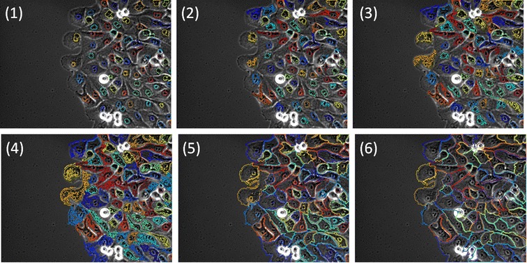
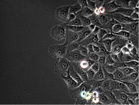
Many cell lines currently used in medical research, such as cancer cells or stem cells, grow in confluent sheets or colonies. The biology of individual cells provide valuable information, thus the separation of touching cells in these microscopy images is critical for counting, identification and measurement of individual cells. Over-segmentation of single cells continues to be a major problem for methods based on morphological watershed due to the high level of noise in microscopy cell images. There is a need for a new segmentation method that is robust over a wide variety of biological images and can accurately separate individual cells even in challenging datasets such as confluent sheets or colonies.

RESULTS

We present a new automated segmentation method called FogBank that accurately separates cells when confluent and touching each other. This technique is successfully applied to phase contrast, bright field, fluorescence microscopy and binary images. The method is based on morphological watershed principles with two new features to improve accuracy and minimize over-segmentation. First, FogBank uses histogram binning to quantize pixel intensities which minimizes the image noise that causes over-segmentation. Second, FogBank uses a geodesic distance mask derived from raw images to detect the shapes of individual cells, in contrast to the more linear cell edges that other watershed-like algorithms produce. We evaluated the segmentation accuracy against manually segmented datasets using two metrics. FogBank achieved segmentation accuracy on the order of 0.75 (1 being a perfect match). We compared our method with other available segmentation techniques in term of achieved performance over the reference data sets. FogBank outperformed all related algorithms. The accuracy has also been visually verified on data sets with 14 cell lines across 3 imaging modalities leading to 876 segmentation evaluation images.

CONCLUSIONS

FogBank produces single cell segmentation from confluent cell sheets with high accuracy. It can be applied to microscopy images of multiple cell lines and a variety of imaging modalities. The code for the segmentation method is available as open-source and includes a Graphical User Interface for user friendly execution.

摘要

背景

目前医学研究中使用的许多细胞系,如癌细胞或干细胞,以汇合的片层或集落形式生长。单个细胞的生物学特性提供了有价值的信息,因此在这些显微镜图像中分离相互接触的细胞对于单个细胞的计数、识别和测量至关重要。由于显微镜细胞图像中的噪声水平较高,基于形态学分水岭的方法中单个细胞的过度分割仍然是一个主要问题。需要一种新的分割方法,该方法在各种生物图像上都具有鲁棒性,并且即使在诸如汇合片层或集落等具有挑战性的数据集中也能准确分离单个细胞。

结果

我们提出了一种名为FogBank的新的自动分割方法,该方法在细胞汇合且相互接触时能准确分离细胞。该技术已成功应用于相差显微镜、明场显微镜、荧光显微镜和二值图像。该方法基于形态学分水岭原理,并具有两个新特性以提高准确性并最小化过度分割。首先,FogBank使用直方图装箱来量化像素强度,从而将导致过度分割的图像噪声降至最低。其次,与其他类似分水岭算法产生的更线性的细胞边缘不同,FogBank使用从原始图像导出的测地距离掩码来检测单个细胞的形状。我们使用两个指标针对手动分割的数据集评估了分割准确性。FogBank实现的分割准确性约为0.75(1表示完美匹配)。我们在参考数据集上的性能方面将我们的方法与其他可用的分割技术进行了比较。FogBank优于所有相关算法。通过对跨越3种成像模式的14个细胞系的数据集进行视觉验证,也验证了其准确性,共生成了876张分割评估图像。

结论

FogBank能从汇合的细胞片中高精度地分割单个细胞。它可应用于多种细胞系的显微镜图像和各种成像模式。该分割方法的代码以开源形式提供,并包括一个图形用户界面以便于用户友好地执行。

https://cdn.ncbi.nlm.nih.gov/pmc/blobs/cfb0/4301455/8205a2776dcf/12859_2014_431_Fig10_HTML.jpg
https://cdn.ncbi.nlm.nih.gov/pmc/blobs/cfb0/4301455/c150fdca9bce/12859_2014_431_Fig1_HTML.jpg
https://cdn.ncbi.nlm.nih.gov/pmc/blobs/cfb0/4301455/2afb5c850c57/12859_2014_431_Fig2_HTML.jpg
https://cdn.ncbi.nlm.nih.gov/pmc/blobs/cfb0/4301455/09211b39a305/12859_2014_431_Fig3_HTML.jpg
https://cdn.ncbi.nlm.nih.gov/pmc/blobs/cfb0/4301455/7560c5d76465/12859_2014_431_Fig4_HTML.jpg
https://cdn.ncbi.nlm.nih.gov/pmc/blobs/cfb0/4301455/0800bd1bd279/12859_2014_431_Fig5_HTML.jpg
https://cdn.ncbi.nlm.nih.gov/pmc/blobs/cfb0/4301455/ab4db13f6867/12859_2014_431_Fig6_HTML.jpg
https://cdn.ncbi.nlm.nih.gov/pmc/blobs/cfb0/4301455/7679d38ae025/12859_2014_431_Fig7_HTML.jpg
https://cdn.ncbi.nlm.nih.gov/pmc/blobs/cfb0/4301455/006f978f7ceb/12859_2014_431_Fig8_HTML.jpg
https://cdn.ncbi.nlm.nih.gov/pmc/blobs/cfb0/4301455/d3b475fa8d92/12859_2014_431_Fig9_HTML.jpg
https://cdn.ncbi.nlm.nih.gov/pmc/blobs/cfb0/4301455/8205a2776dcf/12859_2014_431_Fig10_HTML.jpg

相似文献

1
FogBank: a single cell segmentation across multiple cell lines and image modalities.FogBank:跨多种细胞系和图像模态的单细胞分割
BMC Bioinformatics. 2014 Dec 30;15(1):431. doi: 10.1186/s12859-014-0431-x.
2
Empirical gradient threshold technique for automated segmentation across image modalities and cell lines.用于跨图像模态和细胞系自动分割的经验梯度阈值技术。
J Microsc. 2015 Oct;260(1):86-99. doi: 10.1111/jmi.12269. Epub 2015 Jun 5.
3
CP-CHARM: segmentation-free image classification made accessible.CP-CHARM:实现无分割图像分类。
BMC Bioinformatics. 2016 Jan 27;17:51. doi: 10.1186/s12859-016-0895-y.
4
Segmenting time-lapse phase contrast images of adjacent NIH 3T3 cells.分割相邻 NIH 3T3 细胞的延时相位对比图像。
J Microsc. 2013 Jan;249(1):41-52. doi: 10.1111/j.1365-2818.2012.03678.x. Epub 2012 Nov 5.
5
Robust and automated three-dimensional segmentation of densely packed cell nuclei in different biological specimens with Lines-of-Sight decomposition.利用视线分解对不同生物样本中紧密堆积的细胞核进行稳健且自动化的三维分割。
BMC Bioinformatics. 2015 Jun 8;16:187. doi: 10.1186/s12859-015-0617-x.
6
Comparison of segmentation algorithms for fluorescence microscopy images of cells.细胞荧光显微镜图像分割算法比较。
Cytometry A. 2011 Jul;79(7):545-59. doi: 10.1002/cyto.a.21079. Epub 2011 Jun 14.
7
Cell segmentation in phase contrast microscopy images via semi-supervised classification over optics-related features.基于与光学相关特征的半监督分类的相差显微镜图像中的细胞分割。
Med Image Anal. 2013 Oct;17(7):746-65. doi: 10.1016/j.media.2013.04.004. Epub 2013 Apr 29.
8
CellECT: cell evolution capturing tool.CellECT:细胞进化捕获工具。
BMC Bioinformatics. 2016 Feb 17;17:88. doi: 10.1186/s12859-016-0927-7.
9
Split and Merge Watershed: a two-step method for cell segmentation in fluorescence microscopy images.分裂与合并分水岭算法:一种用于荧光显微镜图像中细胞分割的两步法。
Biomed Signal Process Control. 2019 Aug;53. doi: 10.1016/j.bspc.2019.101575. Epub 2019 Jun 4.
10
Segmentation of yeast cell's bright-field image with an edge-tracing algorithm.利用边缘跟踪算法对酵母细胞明场图像进行分割。
J Biomed Opt. 2018 Nov;23(11):1-7. doi: 10.1117/1.JBO.23.11.116503.

引用本文的文献

1
Label-free live cell recognition and tracking for biological discoveries and translational applications.用于生物学发现和转化应用的无标记活细胞识别与追踪
Npj Imaging. 2024 Oct 7;2(1):41. doi: 10.1038/s44303-024-00046-y.
2
High-volume, label-free imaging for quantifying single-cell dynamics in induced pluripotent stem cell colonies.高通量、无标记成像技术定量分析诱导多能干细胞集落中的单细胞动力学。
PLoS One. 2024 Feb 20;19(2):e0298446. doi: 10.1371/journal.pone.0298446. eCollection 2024.
3
Unbiased image segmentation assessment toolkit for quantitative differentiation of state-of-the-art algorithms and pipelines.

本文引用的文献

1
Empirical gradient threshold technique for automated segmentation across image modalities and cell lines.用于跨图像模态和细胞系自动分割的经验梯度阈值技术。
J Microsc. 2015 Oct;260(1):86-99. doi: 10.1111/jmi.12269. Epub 2015 Jun 5.
2
Cell segmentation by multi-resolution analysis and maximum likelihood estimation (MAMLE).基于多分辨率分析和最大似然估计的细胞分割(MAMLE)。
BMC Bioinformatics. 2013;14 Suppl 10(Suppl 10):S8. doi: 10.1186/1471-2105-14-S10-S8. Epub 2013 Aug 12.
3
An automatic method for robust and fast cell detection in bright field images from high-throughput microscopy.
用于定量区分最先进算法和流程的无偏图像分割评估工具包。
BMC Bioinformatics. 2023 Oct 12;24(1):388. doi: 10.1186/s12859-023-05486-8.
4
Study on segmentation of blasting fragment images from open-pit mine based on U-CARFnet.基于 U-CARFnet 的露天矿爆破块度图像分割研究。
PLoS One. 2023 Sep 14;18(9):e0291115. doi: 10.1371/journal.pone.0291115. eCollection 2023.
5
Dual role of Apolipoprotein D as long-term instructive factor and acute signal conditioning microglial secretory and phagocytic responses.载脂蛋白D作为长期指导因子和急性信号调节小胶质细胞分泌及吞噬反应的双重作用。
Front Cell Neurosci. 2023 Jan 26;17:1112930. doi: 10.3389/fncel.2023.1112930. eCollection 2023.
6
GEMA-An Automatic Segmentation Method for Real-Time Analysis of Mammalian Cell Growth in Microfluidic Devices.GEMA:一种用于微流控装置中哺乳动物细胞生长实时分析的自动分割方法
J Imaging. 2022 Oct 14;8(10):281. doi: 10.3390/jimaging8100281.
7
Ore image segmentation method using U-Net and Res_Unet convolutional networks.基于U-Net和Res_Unet卷积网络的矿石图像分割方法。
RSC Adv. 2020 Mar 4;10(16):9396-9406. doi: 10.1039/c9ra05877j. eCollection 2020 Mar 2.
8
Autocrine signaling can explain the emergence of Allee effects in cancer cell populations.自分泌信号可以解释癌细胞群体中阿利效应的出现。
PLoS Comput Biol. 2022 Mar 3;18(3):e1009844. doi: 10.1371/journal.pcbi.1009844. eCollection 2022 Mar.
9
Robust optical flow algorithm for general single cell segmentation.用于一般单细胞分割的鲁棒光流算法。
PLoS One. 2022 Jan 14;17(1):e0261763. doi: 10.1371/journal.pone.0261763. eCollection 2022.
10
A multi-dimensional, time-lapse, high content screening platform applied to schistosomiasis drug discovery.一种多维、延时、高通量筛选平台,应用于血吸虫病药物发现。
Commun Biol. 2020 Dec 21;3(1):747. doi: 10.1038/s42003-020-01402-5.
一种用于高通量显微镜明场图像中进行稳健快速细胞检测的自动方法。
BMC Bioinformatics. 2013 Oct 4;14:297. doi: 10.1186/1471-2105-14-297.
4
Real-time motion analysis reveals cell directionality as an indicator of breast cancer progression.实时运动分析揭示细胞方向性可作为乳腺癌进展的指标。
PLoS One. 2013;8(3):e58859. doi: 10.1371/journal.pone.0058859. Epub 2013 Mar 19.
5
Segmenting time-lapse phase contrast images of adjacent NIH 3T3 cells.分割相邻 NIH 3T3 细胞的延时相位对比图像。
J Microsc. 2013 Jan;249(1):41-52. doi: 10.1111/j.1365-2818.2012.03678.x. Epub 2012 Nov 5.
6
A fast and robust level set method for image segmentation using fuzzy clustering and lattice Boltzmann method.一种基于模糊聚类和格子玻尔兹曼方法的快速鲁棒图像分割水平集方法。
IEEE Trans Cybern. 2013 Jun;43(3):910-20. doi: 10.1109/TSMCB.2012.2218233. Epub 2012 Oct 11.
7
Automatic segmentation and supervised learning-based selection of nuclei in cancer tissue images.自动分割和基于监督学习的癌症组织图像细胞核选择。
Cytometry A. 2012 Sep;81(9):743-54. doi: 10.1002/cyto.a.22097. Epub 2012 Jul 31.
8
Fiji: an open-source platform for biological-image analysis.斐济:一个用于生物影像分析的开源平台。
Nat Methods. 2012 Jun 28;9(7):676-82. doi: 10.1038/nmeth.2019.
9
Measuring single-cell gene expression dynamics in bacteria using fluorescence time-lapse microscopy.使用荧光延时显微镜测量细菌中单细胞基因表达的动态变化。
Nat Protoc. 2011 Dec 15;7(1):80-8. doi: 10.1038/nprot.2011.432.
10
Interoperability of time series cytometric data: a cross platform approach for modeling tumor heterogeneity.时间序列流式细胞术数据的互操作性:一种用于模拟肿瘤异质性的跨平台方法。
Cytometry A. 2011 Mar;79(3):214-26. doi: 10.1002/cyto.a.21023. Epub 2011 Feb 18.