Department of Internal Medicine, Catholic University of Health and Allied Sciences, P.O Box 1464, Mwanza, Tanzania.
Department of Internal Medicine, Muhimbili University of Health and Allied Sciences, Dar es Salaam, Tanzania.
BMC Cardiovasc Disord. 2021 Oct 9;21(1):485. doi: 10.1186/s12872-021-02297-8.
Left ventricular hypertrophy is a pathophysiological response often due to chronic uncontrolled hypertension. Our primary aim was to investigate the magnitude, correlates and outcomes of left ventricular hypertrophy as a surrogate maker for chronic uncontrolled hypertension in young adults ≤ 45 years with stroke. Our secondary aim was to determine the accuracy of electrocardiography using Sokolow-Lyon and Cornell criteria in detecting left ventricular hypertrophy compared to echocardiography.
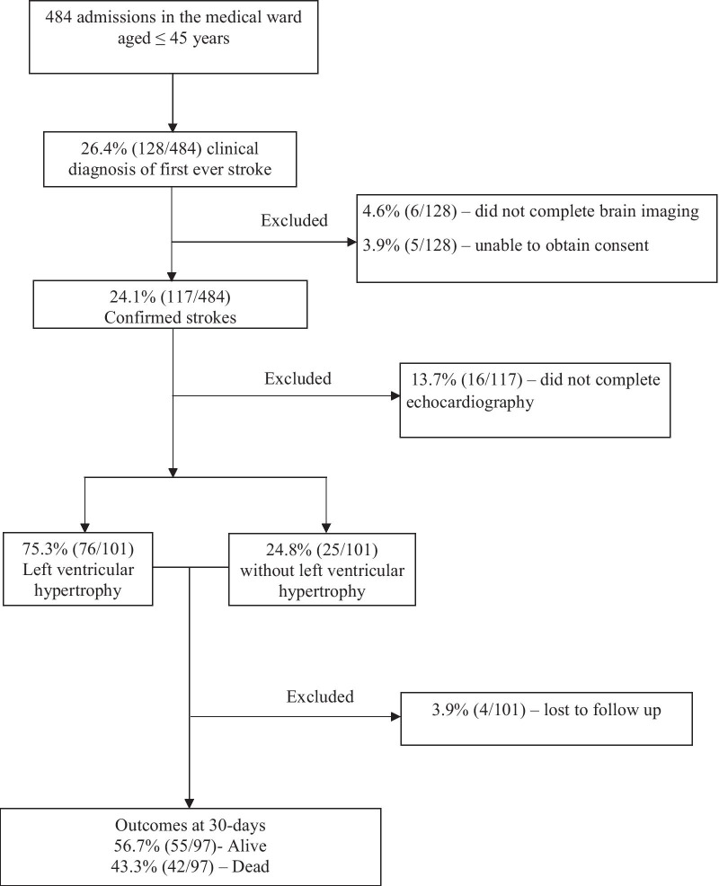
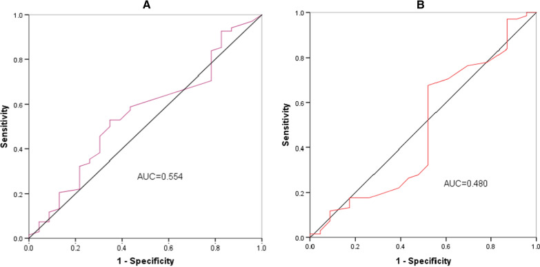
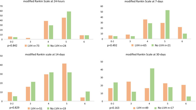
This cohort study recruited young strokes who had undergone brain imaging, electrocardiography and transthoracic echocardiography at baseline. The modified Poisson regression model examined baseline correlates for left ventricular hypertrophy. The National Institute of Health Stroke Scale assessed stroke severity and the modified Rankin Scale assessed outcomes to 30-days. Performance of electrical voltage criterions was estimated using receiver operator characteristics.
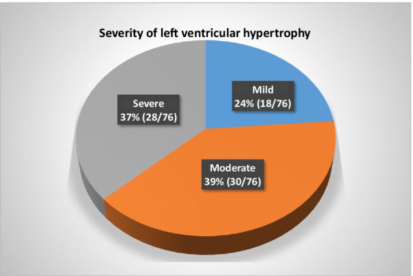
We enrolled 101 stroke participants. Brain imaging revealed ischemic strokes in 60 (59.4%) and those with intracerebral hemorrhage, 33 (86.8%) were localized to the basal ganglia. Left ventricular hypertrophy was present in 76 (75.3%:95%CI 65.7%-83.3%), and 30 (39.5%) and 28 (36.8%) had moderate or severe hypertrophy respectively. Young adults with premorbid or a new diagnosis of hypertension were more likely to have left ventricular hypertrophy, 47 (61.8%), and 26 (34.2%). On multivariable analysis, left ventricular hypertrophy was independently associated with not being on anti-hypertensive medications among hypertensives participants {adjusted risk ratio 1.4 (95%CI:1.04-1.94). The mean National Institute of Health Stroke score was 18 and 30-day mortality was 42 (43.3%). The sensitivity and specificity for Sokolow-Lyon in detecting left ventricular hypertrophy was 27% and 78%, and for Cornell was 32% and 52% respectively.
We identified a high proportion of left ventricular hypertrophy in young adults with stroke associated with chronic undertreated hypertension. While the study methodology does not allow us to determine causation, this association and knowledge of pathophysiological processes supports the notion that chronic hypertension is a major risk factor for young strokes associated with high mortality. Our findings did not support the use of the electrical voltage criteria for detecting left ventricular hypertrophy. We recommend low cost interventions like blood pressure screening and treatment to reduce this burden.
左心室肥厚是一种常见的病理生理反应,通常是由于慢性未控制的高血压引起的。我们的主要目的是研究左心室肥厚的严重程度、相关因素和结局,作为≤45 岁伴有卒中的年轻成人慢性未控制高血压的替代标志物。我们的次要目的是确定心电图 Sokolow-Lyon 和 Cornell 标准与超声心动图相比,在检测左心室肥厚方面的准确性。
这项队列研究招募了在基线时接受了脑部影像学、心电图和经胸超声心动图检查的年轻卒中患者。使用修正泊松回归模型分析左心室肥厚的基线相关因素。国立卫生研究院卒中量表评估卒中严重程度,改良 Rankin 量表评估 30 天的结局。使用接收者操作特征曲线评估电电压标准的性能。
我们共纳入了 101 名卒中患者。脑部影像学显示 60 例(59.4%)为缺血性卒中,其中 33 例(86.8%)为基底节区脑出血。76 例(75.3%:95%CI 65.7%-83.3%)存在左心室肥厚,其中 30 例(39.5%)和 28 例(36.8%)分别为中度或重度肥厚。有高血压前期或新诊断高血压的年轻成年人更有可能存在左心室肥厚,分别为 47 例(61.8%)和 26 例(34.2%)。多变量分析显示,在高血压患者中,未使用抗高血压药物与左心室肥厚独立相关(校正风险比 1.4(95%CI:1.04-1.94))。国立卫生研究院卒中量表的平均评分为 18 分,30 天死亡率为 42 例(43.3%)。Sokolow-Lyon 检测左心室肥厚的敏感性和特异性分别为 27%和 78%,而 Cornell 的敏感性和特异性分别为 32%和 52%。
我们发现,伴有卒中的年轻成年人中存在较高比例的左心室肥厚,与慢性治疗不足的高血压有关。虽然研究方法不能确定因果关系,但这种关联和对病理生理过程的了解支持了慢性高血压是与高死亡率相关的年轻卒中的主要危险因素的观点。我们的研究结果不支持使用电电压标准来检测左心室肥厚。我们建议采取低成本干预措施,如血压筛查和治疗,以减轻这种负担。