Department of Neurology, Graduate School of Medicine, Osaka University, Suita, Osaka, 565-0871, Japan.
Division of Stem Cell Research, Department of Biomedical Research and Innovation, Institute for Clinical Research, National Hospital Organization Osaka National Hospital, Osaka, Osaka, 540-0006, Japan.
Mol Brain. 2021 Oct 11;14(1):149. doi: 10.1186/s13041-021-00851-1.
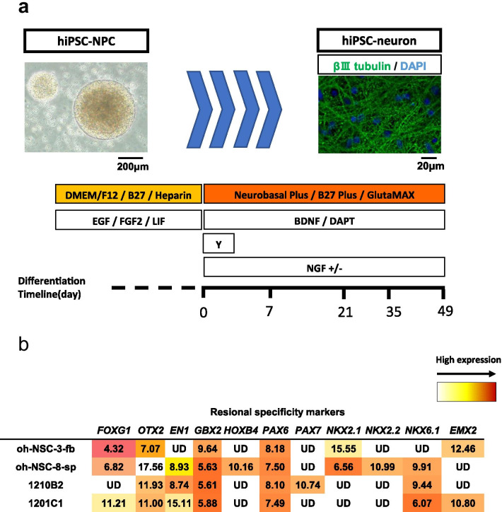
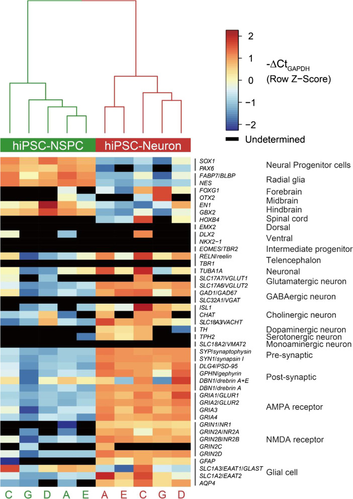
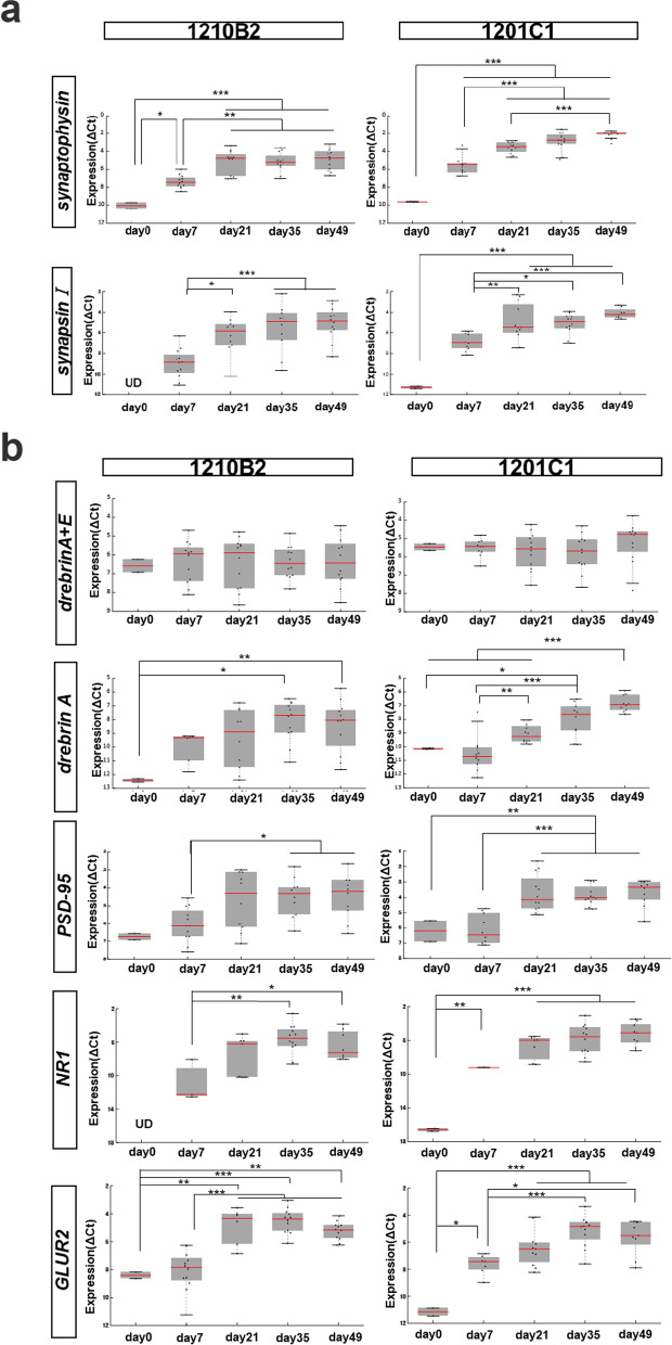
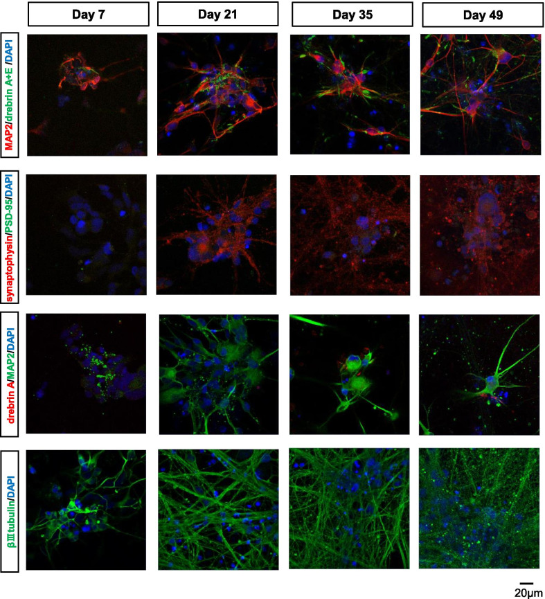
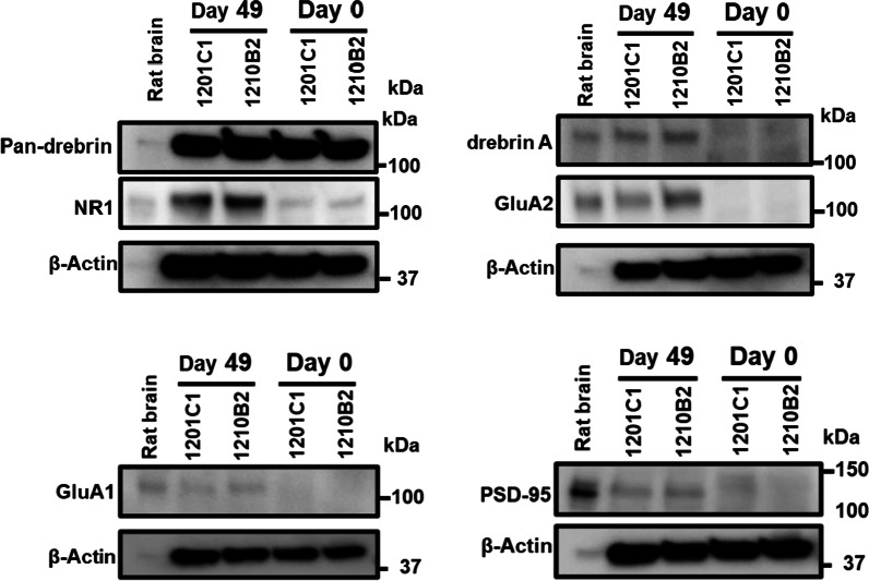
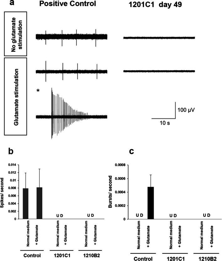
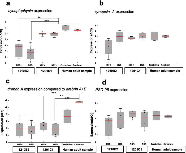
The generation of mature synaptic structures using neurons differentiated from human-induced pluripotent stem cells (hiPSC-neurons) is expected to be applied to physiological studies of synapses in human cells and to pathological studies of diseases that cause abnormal synaptic function. Although it has been reported that synapses themselves change from an immature to a mature state as neurons mature, there are few reports that clearly show when and how human stem cell-derived neurons change to mature synaptic structures. This study was designed to elucidate the synapse formation process of hiPSC-neurons. We propagated hiPSC-derived neural progenitor cells (hiPSC-NPCs) that expressed localized markers of the ventral hindbrain as neurospheres by dual SMAD inhibition and then differentiated them into hiPSC-neurons in vitro. After 49 days of in vitro differentiation, hiPSC-neurons significantly expressed pre- and postsynaptic markers at both the transcript and protein levels. However, the expression of postsynaptic markers was lower than in normal human or normal rat brain tissues, and immunostaining analysis showed that it was relatively modest and was lower than that of presynaptic markers and that its localization in synaptic structures was insufficient. Neurophysiological analysis using a microelectrode array also revealed that no synaptic activity was generated on hiPSC-neurons at 49 days of differentiation. Analysis of subtype markers by immunostaining revealed that most hiPSC-neurons expressed vesicular glutamate transporter 2 (VGLUT2). The presence or absence of NGF, which is required for the survival of cholinergic neurons, had no effect on their cell fractionation. These results suggest that during the synaptogenesis of hiPSC-neurons, the formation of presynaptic structures is not the only requirement for the formation of postsynaptic structures and that the mRNA expression of postsynaptic markers does not correlate with the formation of their mature structures. Technically, we also confirmed a certain level of robustness and reproducibility of our neuronal differentiation method in a multicenter setting, which will be helpful for future research. Synapse formation with mature postsynaptic structures will remain an interesting issue for stem cell-derived neurons, and the present method can be used to obtain early and stable quality neuronal cultures from hiPSC-NPCs.
利用人诱导多能干细胞(hiPSC)分化而来的神经元生成成熟的突触结构,有望应用于人类细胞中突触的生理学研究,以及导致突触功能异常的疾病的病理学研究。虽然已经有报道称,随着神经元的成熟,突触本身会从不成熟状态转变为成熟状态,但很少有报道清楚地表明人类干细胞来源的神经元何时以及如何转变为成熟的突触结构。本研究旨在阐明 hiPSC 神经元的突触形成过程。我们通过双重 SMAD 抑制增殖表达腹侧后脑局部标志物的 hiPSC 衍生神经前体细胞(hiPSC-NPC),然后在体外将其分化为 hiPSC 神经元。在体外分化 49 天后,hiPSC 神经元在转录和蛋白水平上均显著表达了前突触和后突触标志物。然而,后突触标志物的表达低于正常人类或正常大鼠脑组织,免疫染色分析表明其相对较低,低于前突触标志物,且其在突触结构中的定位不足。使用微电极阵列的神经生理学分析也表明,在分化 49 天时,hiPSC 神经元上没有产生突触活性。免疫染色分析的亚型标志物分析表明,大多数 hiPSC 神经元表达囊泡谷氨酸转运体 2(VGLUT2)。存在或不存在神经营养因子(NGF),它是胆碱能神经元存活所必需的,对其细胞分离没有影响。这些结果表明,在 hiPSC 神经元的突触发生过程中,前突触结构的形成不仅仅是后突触结构形成的唯一要求,后突触标志物的 mRNA 表达与它们成熟结构的形成无关。从技术上讲,我们还在多中心环境中证实了我们的神经元分化方法具有一定的稳健性和可重复性,这将有助于未来的研究。具有成熟后突触结构的突触形成仍然是干细胞源性神经元的一个有趣问题,本方法可用于从 hiPSC-NPC 获得早期和稳定质量的神经元培养物。