• 文献检索
  • 文档翻译
  • 深度研究
  • 学术资讯
  • Suppr Zotero 插件Zotero 插件
  • 邀请有礼
  • 套餐&价格
  • 历史记录
应用&插件
Suppr Zotero 插件Zotero 插件浏览器插件Mac 客户端Windows 客户端微信小程序
定价
高级版会员购买积分包购买API积分包
服务
文献检索文档翻译深度研究API 文档MCP 服务
关于我们
关于 Suppr公司介绍联系我们用户协议隐私条款
关注我们

Suppr 超能文献

核心技术专利:CN118964589B侵权必究
粤ICP备2023148730 号-1Suppr @ 2026

文献检索

告别复杂PubMed语法,用中文像聊天一样搜索,搜遍4000万医学文献。AI智能推荐,让科研检索更轻松。

立即免费搜索

文件翻译

保留排版,准确专业,支持PDF/Word/PPT等文件格式,支持 12+语言互译。

免费翻译文档

深度研究

AI帮你快速写综述,25分钟生成高质量综述,智能提取关键信息,辅助科研写作。

立即免费体验

转化生长因子-β受体II对出生后牙槽骨形成过程中成骨祖细胞的增殖和分化至关重要。

TGF-Beta Receptor II Is Critical for Osteogenic Progenitor Cell Proliferation and Differentiation During Postnatal Alveolar Bone Formation.

作者信息

Xu Chunmei, Xie Xudong, Zhao Hu, Wu Yafei, Wang Jun, Feng Jian Q

机构信息

State Key Laboratory of Oral Diseases, National Clinical Research Center for Oral Diseases, Department of Periodontics, West China Hospital of Stomatology, Sichuan University, Chengdu, China.

Department of Biomedical Sciences, College of Dentistry, Texas A&M University, Dallas, TX, United States.

出版信息

Front Physiol. 2021 Sep 24;12:721775. doi: 10.3389/fphys.2021.721775. eCollection 2021.

DOI:10.3389/fphys.2021.721775
PMID:34630143
原文链接:https://pmc.ncbi.nlm.nih.gov/articles/PMC8497707/
Abstract

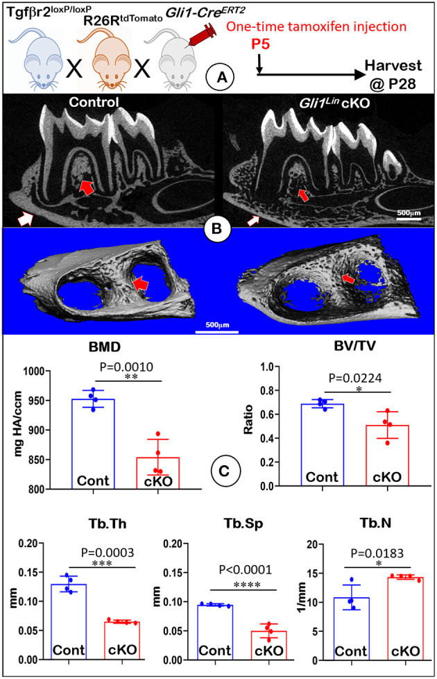
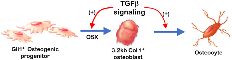
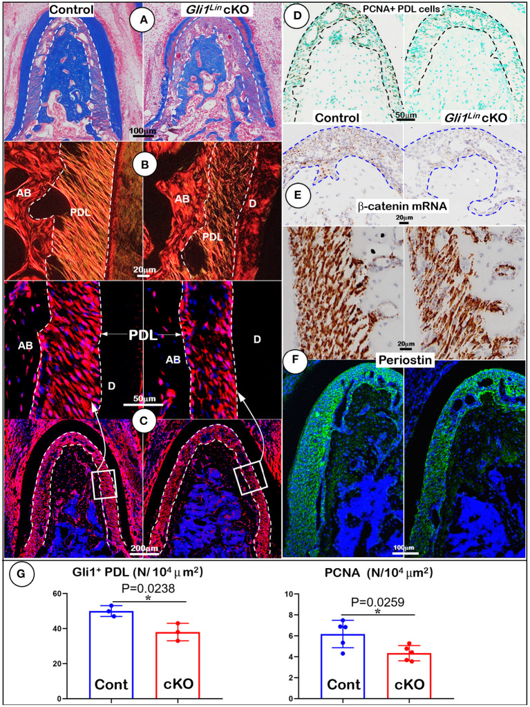
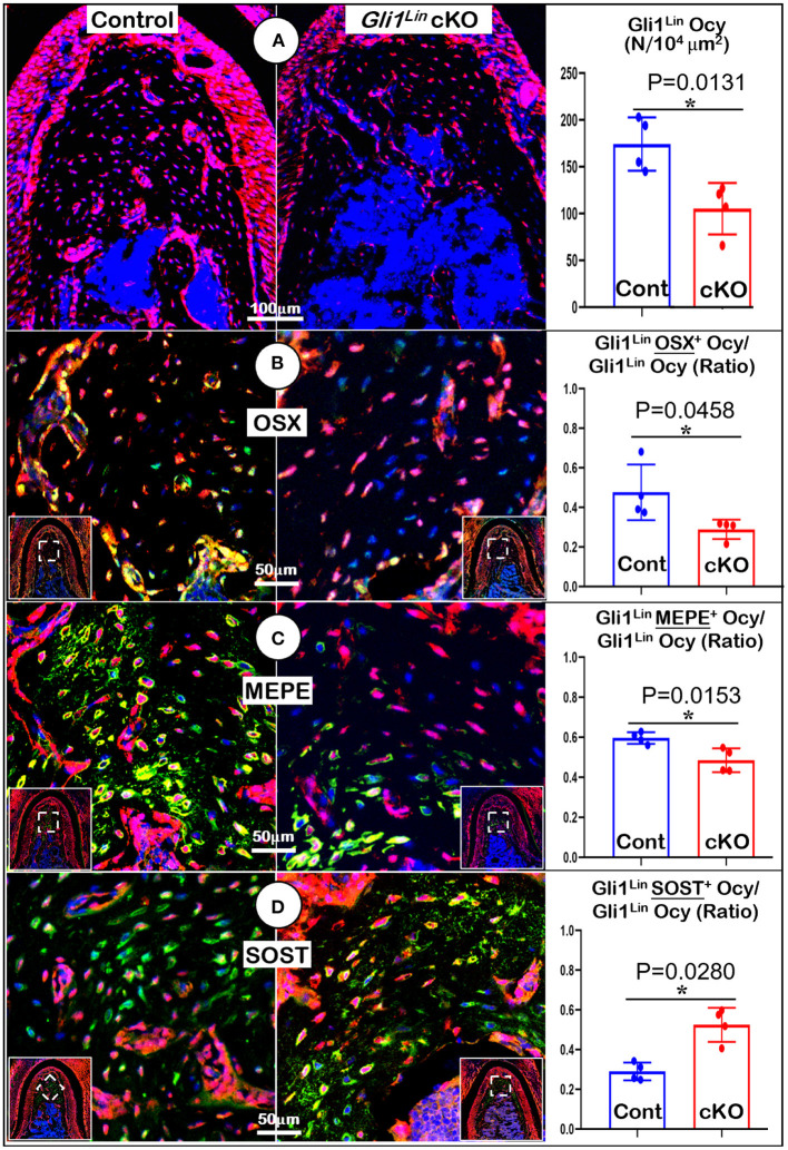
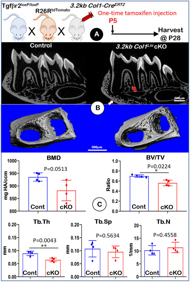
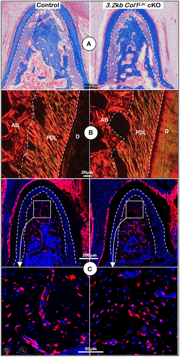
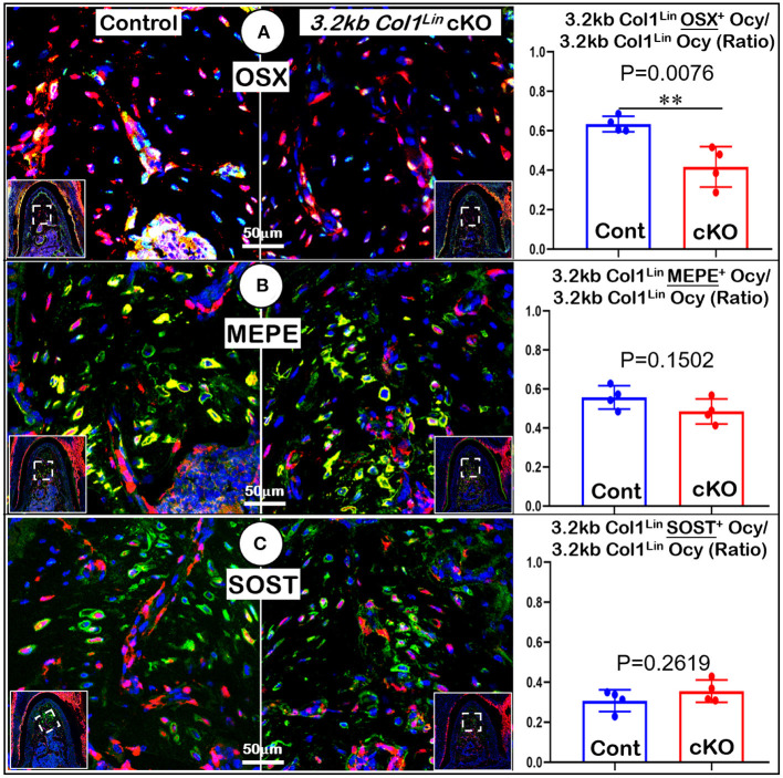
Transforming growth factor beta (TGFβ) signaling plays an important role during osteogenesis. However, most research in this area focuses on cortical and trabecular bone, whereas alveolar bone is largely overlooked. To address the role of TGFβR2 (the key receptor for TGFβ signaling) during postnatal alveolar bone development, we conditionally deleted β in early mesenchymal progenitors by crossing ; β ; mice (named early cKO) or in osteoblasts by crossing β mice (named late cKO). Both cKO lines were induced at postnatal day 5 (P5) and mice were harvested at P28. Compared to the control littermates, early cKO mice exhibited significant reduction in alveolar bone mass and bone mineral density, with drastic defects in the periodontal ligament (PDL); conversely, the late cKO mice displayed very minor changes in alveolar bone. Mechanism studies showed a significant reduction in PCNA+ PDL cell numbers and OSX+ alveolar bone cell numbers, as well as disorganized PDL fibers with a great reduction in periostin (the most abundant extracellular matrix protein) on both mRNA and protein levels. We also showed a drastic reduction in β-catenin in the early cKO PDL and a great increase in SOST (a potent inhibitor of Wnt signaling). Based on these findings, we conclude that TGFβ signaling plays critical roles during early alveolar bone formation via the promotion of PDL mesenchymal progenitor proliferation and differentiation mechanisms.

摘要

转化生长因子β(TGFβ)信号通路在骨生成过程中发挥着重要作用。然而,该领域的大多数研究都集中在皮质骨和小梁骨,而牙槽骨在很大程度上被忽视了。为了研究TGFβR2(TGFβ信号通路的关键受体)在出生后牙槽骨发育中的作用,我们通过杂交;β;;小鼠(命名为早期cKO)在早期间充质祖细胞中条件性删除β,或通过杂交β小鼠(命名为晚期cKO)在成骨细胞中条件性删除β。两个cKO品系均在出生后第5天(P5)诱导,小鼠在P28处死取材。与对照同窝小鼠相比,早期cKO小鼠的牙槽骨量和骨矿物质密度显著降低,牙周韧带(PDL)出现严重缺陷;相反,晚期cKO小鼠的牙槽骨变化非常小。机制研究表明,PCNA+ PDL细胞数量和OSX+牙槽骨细胞数量显著减少,PDL纤维排列紊乱,骨膜蛋白(最丰富的细胞外基质蛋白)在mRNA和蛋白质水平上均大幅减少。我们还发现早期cKO的PDL中β-连环蛋白大幅减少,而sost(一种有效的Wnt信号抑制剂)大幅增加。基于这些发现,我们得出结论,TGFβ信号通路通过促进PDL间充质祖细胞增殖和分化机制,在早期牙槽骨形成过程中发挥关键作用。

https://cdn.ncbi.nlm.nih.gov/pmc/blobs/11d8/8497707/4a2dd3113eff/fphys-12-721775-g0007.jpg
https://cdn.ncbi.nlm.nih.gov/pmc/blobs/11d8/8497707/284618ec3f80/fphys-12-721775-g0001.jpg
https://cdn.ncbi.nlm.nih.gov/pmc/blobs/11d8/8497707/2a06f4fa29ba/fphys-12-721775-g0002.jpg
https://cdn.ncbi.nlm.nih.gov/pmc/blobs/11d8/8497707/ce65dab3e837/fphys-12-721775-g0003.jpg
https://cdn.ncbi.nlm.nih.gov/pmc/blobs/11d8/8497707/8d1ac5a0a4d3/fphys-12-721775-g0004.jpg
https://cdn.ncbi.nlm.nih.gov/pmc/blobs/11d8/8497707/13c1efdea5f9/fphys-12-721775-g0005.jpg
https://cdn.ncbi.nlm.nih.gov/pmc/blobs/11d8/8497707/b52b63758788/fphys-12-721775-g0006.jpg
https://cdn.ncbi.nlm.nih.gov/pmc/blobs/11d8/8497707/4a2dd3113eff/fphys-12-721775-g0007.jpg

相似文献

1
TGF-Beta Receptor II Is Critical for Osteogenic Progenitor Cell Proliferation and Differentiation During Postnatal Alveolar Bone Formation.转化生长因子-β受体II对出生后牙槽骨形成过程中成骨祖细胞的增殖和分化至关重要。
Front Physiol. 2021 Sep 24;12:721775. doi: 10.3389/fphys.2021.721775. eCollection 2021.
2
Axin2+ PDL Cells Directly Contribute to New Alveolar Bone Formation in Response to Orthodontic Tension Force.Axin2+ PDL 细胞直接促进正畸张力刺激下新牙槽骨形成。
J Dent Res. 2022 Jun;101(6):695-703. doi: 10.1177/00220345211062585. Epub 2022 Jan 9.
3
Bone formation ability of Gli1 cells in the periodontal ligament after tooth extraction.拔牙后牙周膜中 Gli1 细胞的成骨能力。
Bone. 2023 Aug;173:116786. doi: 10.1016/j.bone.2023.116786. Epub 2023 May 8.
4
Osteoblast differentiation of Gli1⁺ cells via Wnt and BMP signaling pathways during orthodontic tooth movement.Gli1⁺ 细胞通过 Wnt 和 BMP 信号通路在正畸牙齿移动过程中的成骨细胞分化。
J Oral Biosci. 2024 Jun;66(2):373-380. doi: 10.1016/j.job.2024.03.004. Epub 2024 Mar 16.
5
Disruption of Kif3a in osteoblasts results in defective bone formation and osteopenia.破骨细胞中 Kif3a 的缺失导致骨形成缺陷和骨质疏松症。
J Cell Sci. 2012 Apr 15;125(Pt 8):1945-57. doi: 10.1242/jcs.095893. Epub 2012 Feb 22.
6
ACVR1 is essential for periodontium development and promotes alveolar bone formation.ACVR1 对于牙周组织的发育是必不可少的,并且促进牙槽骨的形成。
Arch Oral Biol. 2018 Nov;95:108-117. doi: 10.1016/j.archoralbio.2018.07.019. Epub 2018 Jul 31.
7
LRP1-deficient leptin receptor-positive cells in periodontal ligament tissue reduce alveolar bone mass by inhibiting bone formation.LRP1 缺乏的瘦素受体阳性细胞通过抑制骨形成减少牙周韧带组织中的牙槽骨量。
Arch Oral Biol. 2024 Feb;158:105853. doi: 10.1016/j.archoralbio.2023.105853. Epub 2023 Nov 23.
8
Differentiation ability of Gli1 cells during orthodontic tooth movement.Gli1 细胞在正畸牙齿移动过程中的分化能力。
Bone. 2023 Jan;166:116609. doi: 10.1016/j.bone.2022.116609. Epub 2022 Nov 9.
9
A Biphasic Feature of Gli1-Mesenchymal Progenitors during Cementogenesis That Is Positively Controlled by Wnt/β-Catenin Signaling.成骨过程中Gli1 间质祖细胞的双相特征受 Wnt/β-连环蛋白信号的正向调控。
J Dent Res. 2021 Oct;100(11):1289-1298. doi: 10.1177/00220345211007429. Epub 2021 Apr 14.
10
Conditional disruption of the gene in chondrocytes during early postnatal growth impairs secondary ossification in the mouse tibial epiphysis.出生后早期生长过程中软骨细胞中该基因的条件性破坏会损害小鼠胫骨骺的次级骨化。
Bone Res. 2019 Aug 5;7:24. doi: 10.1038/s41413-019-0064-9. eCollection 2019.

引用本文的文献

1
Bone or Tooth dentin: The TGF-β signaling is the key.骨或牙本质:TGF-β 信号是关键。
Int J Biol Sci. 2024 Jun 24;20(9):3557-3569. doi: 10.7150/ijbs.97206. eCollection 2024.
2
A role for TGFβ signaling in Gli1+ tendon and enthesis cells.TGFβ 信号在 Gli1+ 肌腱和腱止点细胞中的作用。
FASEB J. 2024 Mar 31;38(6):e23568. doi: 10.1096/fj.202301452R.
3
The Role of Gli1 Mesenchymal Stem Cells in Osteogenesis of Craniofacial Bone.Gli1间充质干细胞在颅面骨成骨中的作用

本文引用的文献

1
Skeletal Deformities in Osterix-Cre;Tgfbr2 Mice May Cause Postnatal Death.Osterix-Cre;Tgfbr2 小鼠的骨骼畸形可能导致出生后死亡。
Genes (Basel). 2021 Jun 25;12(7):975. doi: 10.3390/genes12070975.
2
Alveolar Bone Marrow Gli1+ Stem Cells Support Implant Osseointegration.肺泡骨髓Gli1+ 干细胞支持种植体骨整合。
J Dent Res. 2022 Jan;101(1):73-82. doi: 10.1177/00220345211013722. Epub 2021 May 19.
3
The identification of critical time windows of postnatal root elongation in response to Wnt/β-catenin signaling.鉴定对 Wnt/β-连环蛋白信号反应的出生后根伸长的关键时间窗。
Biomolecules. 2023 Sep 5;13(9):1351. doi: 10.3390/biom13091351.
4
Isolation, Identification, and Characterization of Bioactive Peptides in Human Bone Cells from Tortoiseshell and Deer Antler Gelatin.从龟甲和鹿角明胶中分离、鉴定和表征人成骨细胞中的生物活性肽。
Int J Mol Sci. 2023 Jan 16;24(2):1759. doi: 10.3390/ijms24021759.
5
Intermittent compressive force regulates dentin matrix protein 1 expression in human periodontal ligament stem cells.间歇性压缩力调节人牙周膜干细胞中牙本质基质蛋白1的表达。
J Dent Sci. 2023 Jan;18(1):105-111. doi: 10.1016/j.jds.2022.07.003. Epub 2022 Jul 15.
6
MicroRNA-223 negatively regulates the osteogenic differentiation of periodontal ligament derived cells by directly targeting growth factor receptors.微小 RNA-223 通过直接靶向生长因子受体负调控牙周膜来源细胞的成骨分化。
J Transl Med. 2022 Oct 11;20(1):465. doi: 10.1186/s12967-022-03676-1.
7
Mechanisms of bone remodeling and therapeutic strategies in chronic apical periodontitis.慢性根尖周炎中骨改建的机制与治疗策略。
Front Cell Infect Microbiol. 2022 Jul 22;12:908859. doi: 10.3389/fcimb.2022.908859. eCollection 2022.
Oral Dis. 2022 Mar;28(2):442-451. doi: 10.1111/odi.13753. Epub 2021 Jan 16.
4
TGF-β/Smad2 signalling regulates enchondral bone formation of Gli1 periosteal cells during fracture healing.TGF-β/Smad2 信号通路调控 Gli1 骨膜细胞在骨折愈合过程中的软骨内骨形成。
Cell Prolif. 2020 Nov;53(11):e12904. doi: 10.1111/cpr.12904. Epub 2020 Sep 30.
5
Stem cell properties of Gli1-positive cells in the periodontal ligament.牙周膜Gli1 阳性细胞的干细胞特性。
J Oral Biosci. 2020 Dec;62(4):299-305. doi: 10.1016/j.job.2020.08.002. Epub 2020 Aug 31.
6
Gli1+ Periodontium Stem Cells Are Regulated by Osteocytes and Occlusal Force.Gli1+ 牙周干细胞受破骨细胞和成牙骨质细胞以及咬合压力的调节。
Dev Cell. 2020 Sep 14;54(5):639-654.e6. doi: 10.1016/j.devcel.2020.06.006. Epub 2020 Jul 10.
7
Mechanosensing by Gli1 cells contributes to the orthodontic force-induced bone remodelling.Gli1 细胞的机械感知有助于正畸力诱导的骨重塑。
Cell Prolif. 2020 May;53(5):e12810. doi: 10.1111/cpr.12810. Epub 2020 Apr 23.
8
Axin2-Mesenchymal PDL Cells, Instead of K14 Epithelial Cells, Play a Key Role in Rapid Cementum Growth.Axin2 间质 PDLCs,而非 K14 上皮细胞,在快速牙骨质生长中发挥关键作用。
J Dent Res. 2019 Oct;98(11):1262-1270. doi: 10.1177/0022034519871021. Epub 2019 Aug 27.
9
TGF-β Signaling Plays an Essential Role in the Lineage Specification of Mesenchymal Stem/Progenitor Cells in Fetal Bone Marrow.TGF-β 信号在胎儿骨髓间充质干细胞/祖细胞的谱系特化中发挥重要作用。
Stem Cell Reports. 2019 Jul 9;13(1):48-60. doi: 10.1016/j.stemcr.2019.05.017. Epub 2019 Jun 13.
10
Runx2 regulates cranial suture closure by inducing hedgehog, Fgf, Wnt and Pthlh signaling pathway gene expressions in suture mesenchymal cells.Runx2 通过诱导颅缝间质细胞中 hedgehog、Fgf、Wnt 和 Pthlh 信号通路基因的表达来调节颅缝闭合。
Hum Mol Genet. 2019 Mar 15;28(6):896-911. doi: 10.1093/hmg/ddy386.