Department of Neurosurgery, the First Affiliated Hospital of Soochow University, No188, Shizi Street, Suzhou, 215007, Jiangsu, China.
Department of Neurosurgery, Dushu Lake Hospital Affiliated of Soochow University, Suzhou, 215124, Jiangsu, China.
Cell Death Dis. 2021 Oct 11;12(10):927. doi: 10.1038/s41419-021-04225-2.
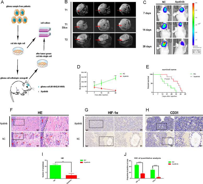
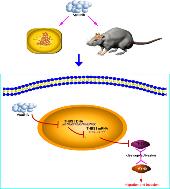
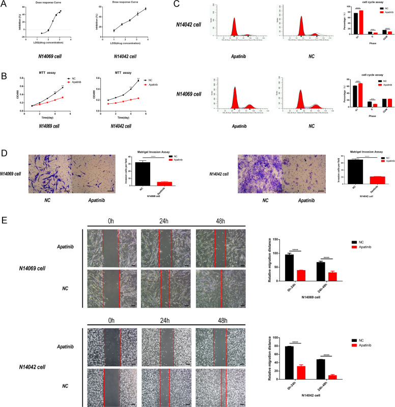
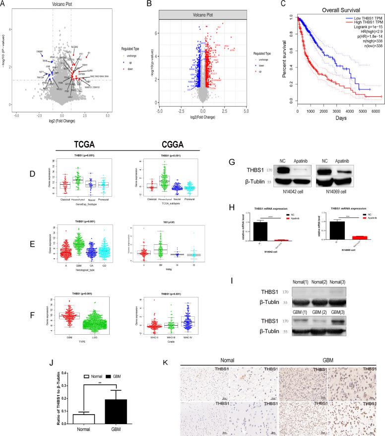
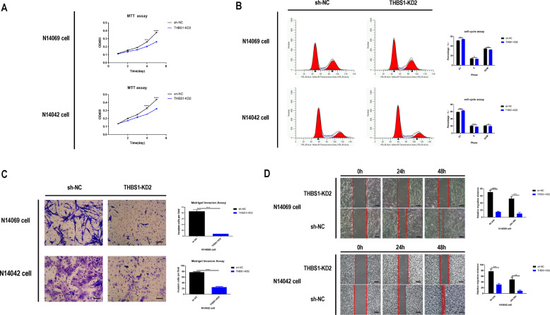
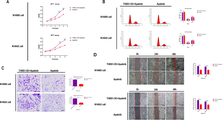
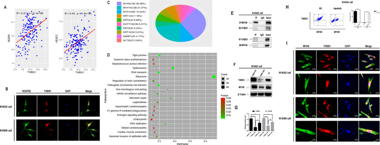
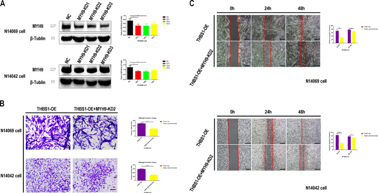
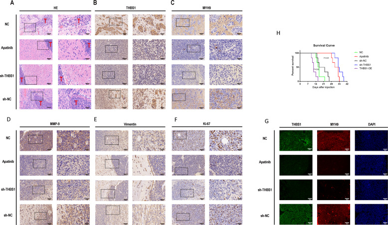
We determined the antitumor mechanism of apatinib in glioma using a patient-derived orthotopic xenograft (PDOX) glioma mouse model and glioblastoma (GBM) cell lines. The PDOX mouse model was established using tumor tissues from two glioma patients via single-cell injections. Sixteen mice were successfully modeled and randomly divided into two equal groups (n = 8/group): apatinib and normal control. Survival analysis and in vivo imaging was performed to determine the effect of apatinib on glioma proliferation in vivo. Candidate genes in GBM cells that may be affected by apatinib treatment were screened using RNA-sequencing coupled with quantitative mass spectrometry, data mining of The Cancer Genome Atlas, and Chinese Glioma Genome Atlas databases, and immunohistochemistry analysis of clinical high-grade glioma pathology samples. Quantitative reverse transcription-polymerase chain reaction (qPCR), western blotting, and co-immunoprecipitation (co-IP) were performed to assess gene expression and the apatinib-mediated effect on glioma cell malignancy. Apatinib inhibited the proliferation and malignancy of glioma cells in vivo and in vitro. Thrombospondin 1 (THBS1) was identified as a potential target of apatinib that lead to inhibited glioma cell proliferation. Apatinib-mediated THBS1 downregulation in glioma cells was confirmed by qPCR and western blotting. Co-IP and mass spectrometry analysis revealed that THBS1 could interact with myosin heavy chain 9 (MYH9) in glioma cells. Simultaneous THBS1 overexpression and MYH9 knockdown suppressed glioma cell invasion and migration. These data suggest that apatinib targets THBS1 in glioma cells, potentially via MYH9, to inhibit glioma cell malignancy and may provide novel targets for glioma therapy.
我们使用患者来源的原位异种移植(PDOX)脑胶质瘤小鼠模型和脑胶质瘤(GBM)细胞系来确定阿帕替尼在脑胶质瘤中的抗肿瘤机制。通过单细胞注射,使用来自两名脑胶质瘤患者的肿瘤组织建立了 PDOX 小鼠模型。成功建立了 16 只小鼠模型,并将其随机分为两组(每组 n = 8):阿帕替尼组和正常对照组。进行生存分析和体内成像,以确定阿帕替尼对体内脑胶质瘤增殖的影响。通过 RNA 测序结合定量质谱、癌症基因组图谱和中国脑胶质瘤基因组图谱数据库的数据挖掘,筛选出可能受阿帕替尼治疗影响的 GBM 细胞中的候选基因,并对临床高级别脑胶质瘤病理样本进行免疫组织化学分析。采用定量逆转录聚合酶链反应(qPCR)、蛋白质印迹和免疫共沉淀(co-IP)来评估基因表达以及阿帕替尼对脑胶质瘤细胞恶性程度的影响。阿帕替尼在体内和体外抑制脑胶质瘤细胞的增殖和恶性程度。血小板反应蛋白 1(THBS1)被鉴定为阿帕替尼的一个潜在靶点,可导致脑胶质瘤细胞增殖受到抑制。qPCR 和蛋白质印迹证实了阿帕替尼介导的脑胶质瘤细胞中 THBS1 的下调。co-IP 和质谱分析表明,THBS1 可与肌球蛋白重链 9(MYH9)在脑胶质瘤细胞中相互作用。同时过表达 THBS1 和敲低 MYH9 可抑制脑胶质瘤细胞侵袭和迁移。这些数据表明,阿帕替尼在脑胶质瘤细胞中靶向 THBS1,可能通过 MYH9 抑制脑胶质瘤细胞的恶性程度,并可能为脑胶质瘤治疗提供新的靶点。