• 文献检索
  • 文档翻译
  • 深度研究
  • 学术资讯
  • Suppr Zotero 插件Zotero 插件
  • 邀请有礼
  • 套餐&价格
  • 历史记录
应用&插件
Suppr Zotero 插件Zotero 插件浏览器插件Mac 客户端Windows 客户端微信小程序
定价
高级版会员购买积分包购买API积分包
服务
文献检索文档翻译深度研究API 文档MCP 服务
关于我们
关于 Suppr公司介绍联系我们用户协议隐私条款
关注我们

Suppr 超能文献

核心技术专利:CN118964589B侵权必究
粤ICP备2023148730 号-1Suppr @ 2026

文献检索

告别复杂PubMed语法,用中文像聊天一样搜索,搜遍4000万医学文献。AI智能推荐,让科研检索更轻松。

立即免费搜索

文件翻译

保留排版,准确专业,支持PDF/Word/PPT等文件格式,支持 12+语言互译。

免费翻译文档

深度研究

AI帮你快速写综述,25分钟生成高质量综述,智能提取关键信息,辅助科研写作。

立即免费体验

IRAK1 依赖性 Regnase-1-14-3-3 复合物形成控制 Regnase-1 介导的 mRNA 降解。

IRAK1-dependent Regnase-1-14-3-3 complex formation controls Regnase-1-mediated mRNA decay.

机构信息

Department of Medical Chemistry, Graduate School of Medicine, Kyoto University, Kyoto, Japan.

Graduate School of Biostudies, Kyoto University, Kyoto, Japan.

出版信息

Elife. 2021 Oct 12;10:e71966. doi: 10.7554/eLife.71966.

DOI:10.7554/eLife.71966
PMID:34636324
原文链接:https://pmc.ncbi.nlm.nih.gov/articles/PMC8553338/
Abstract

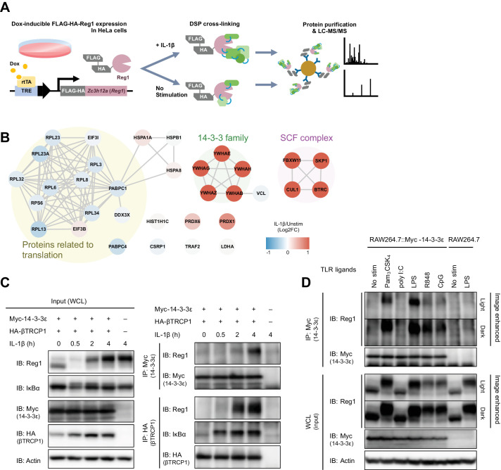
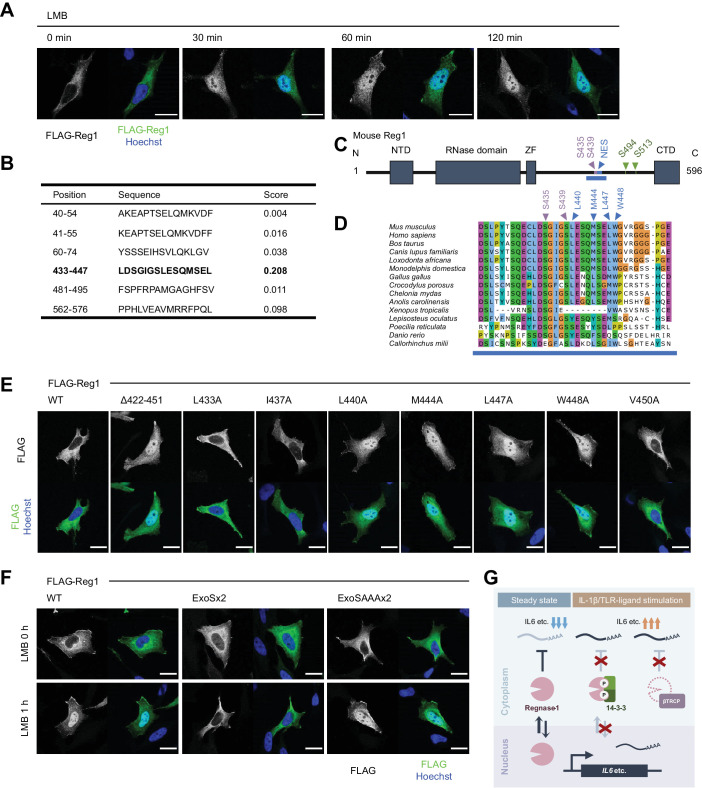
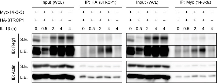
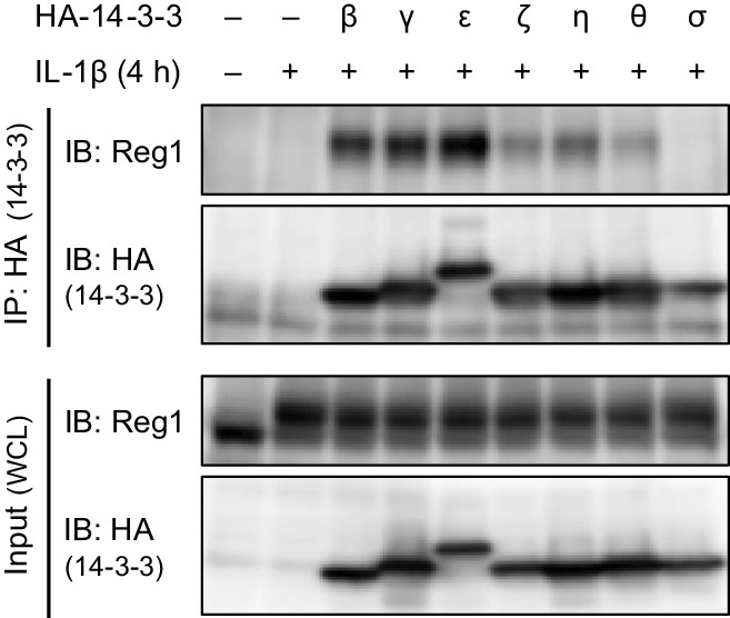
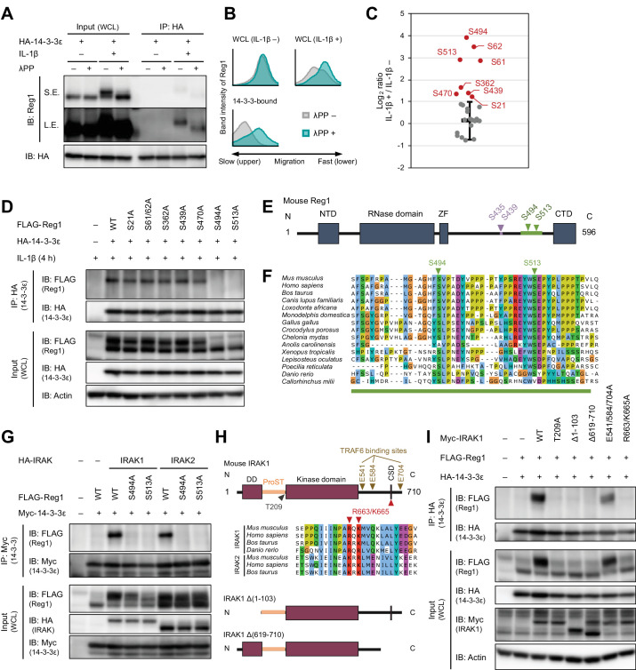
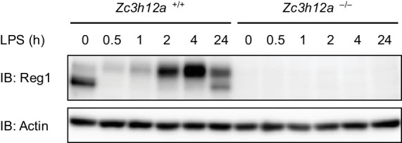
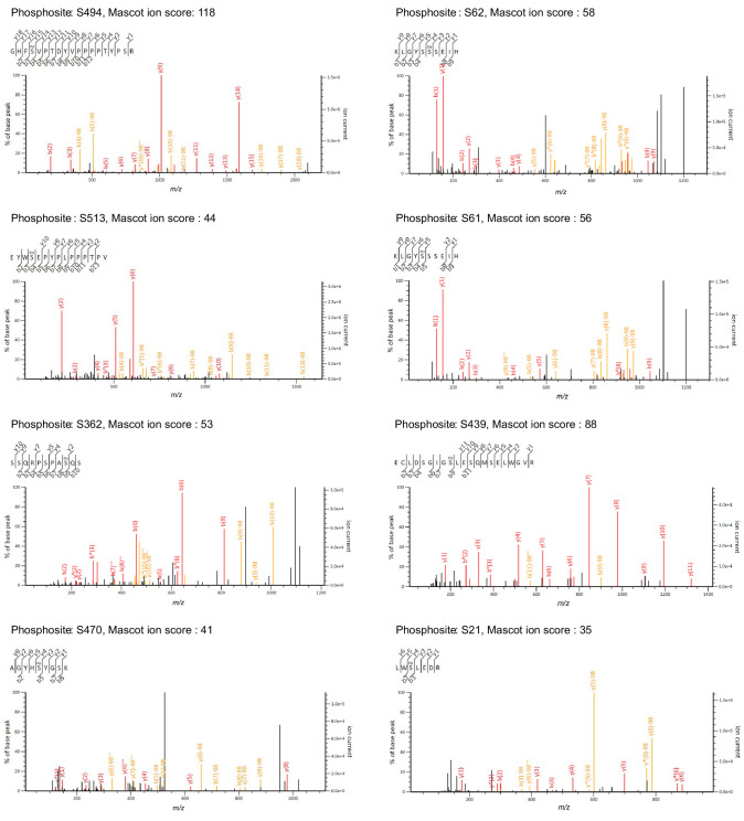
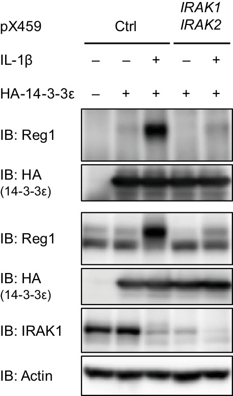
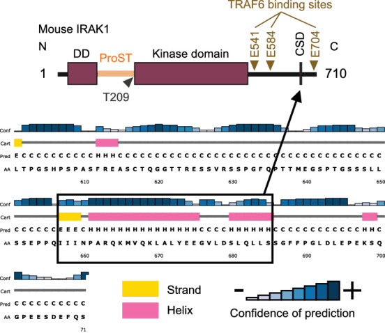
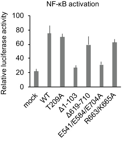
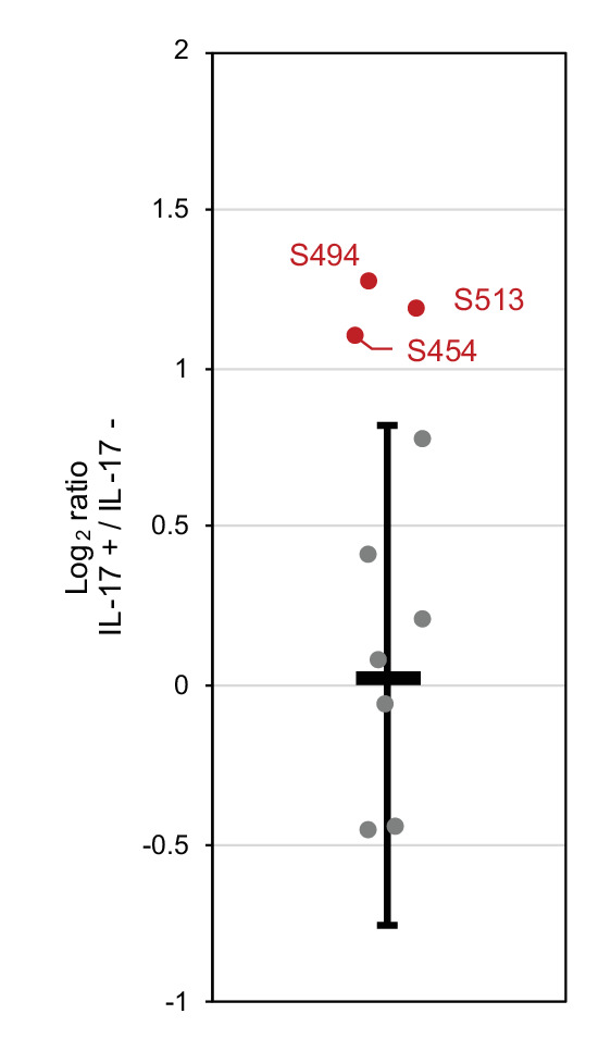
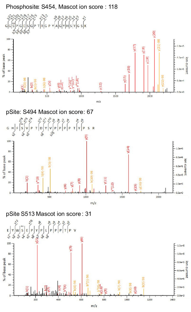
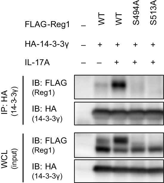
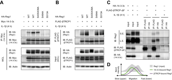
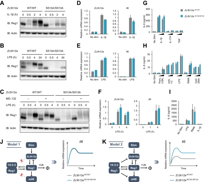
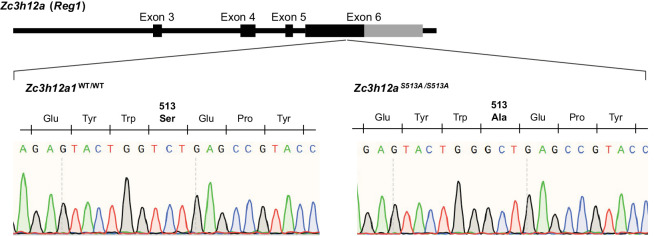
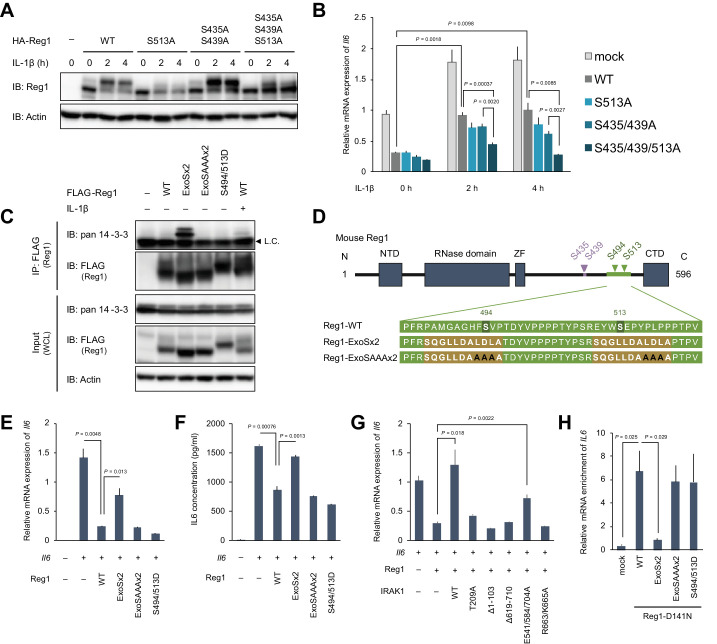
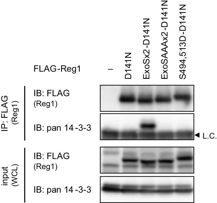
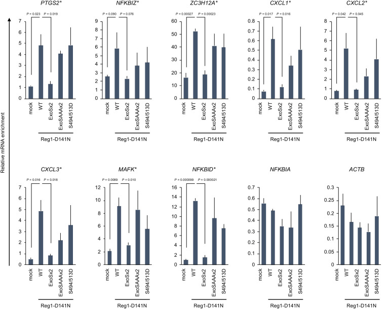
Regnase-1 is an endoribonuclease crucial for controlling inflammation by degrading mRNAs encoding cytokines and inflammatory mediators in mammals. However, it is unclear how Regnase-1-mediated mRNA decay is controlled in interleukin (IL)-1β- or Toll-like receptor (TLR) ligand-stimulated cells. Here, by analyzing the Regnase-1 interactome, we found that IL-1β or TLR stimulus dynamically induced the formation of Regnase-1-β-transducin repeat-containing protein (βTRCP) complex. Importantly, we also uncovered a novel interaction between Regnase-1 and 14-3-3 in both mouse and human cells. In IL-1R/TLR-stimulated cells, the Regnase-1-14-3-3 interaction is mediated by IRAK1 through a previously uncharacterized C-terminal structural domain. Phosphorylation of Regnase-1 at S494 and S513 is critical for Regnase-1-14-3-3 interaction, while a different set of phosphorylation sites of Regnase-1 is known to be required for the recognition by βTRCP and proteasome-mediated degradation. We found that Regnase-1-14-3-3 and Regnase-1-βTRCP interactions are not sequential events. Rather, 14-3-3 protects Regnase-1 from βTRCP-mediated degradation. On the other hand, 14-3-3 abolishes Regnase-1-mediated mRNA decay by inhibiting Regnase-1-mRNA association. In addition, nuclear-cytoplasmic shuttling of Regnase-1 is abrogated by 14-3-3 interaction. Taken together, the results suggest that a novel inflammation-induced interaction of 14-3-3 with Regnase-1 stabilizes inflammatory mRNAs by sequestering Regnase-1 in the cytoplasm to prevent mRNA recognition.

摘要

Regnase-1 是一种内切核酸酶,对于哺乳动物通过降解编码细胞因子和炎症介质的 mRNA 来控制炎症至关重要。然而,Regnase-1 介导的 mRNA 降解如何在白细胞介素 (IL)-1β 或 Toll 样受体 (TLR) 配体刺激的细胞中受到控制尚不清楚。在这里,通过分析 Regnase-1 相互作用组,我们发现 IL-1β 或 TLR 刺激动态诱导形成 Regnase-1-β-转导重复蛋白 (βTRCP) 复合物。重要的是,我们还在小鼠和人类细胞中发现了 Regnase-1 和 14-3-3 之间的一种新相互作用。在 IL-1R/TLR 刺激的细胞中,Regnase-1-14-3-3 相互作用是通过 IRAK1 通过一个以前未表征的 C 末端结构域介导的。Regnase-1 的 S494 和 S513 磷酸化对于 Regnase-1-14-3-3 相互作用至关重要,而 Regnase-1 的另一组磷酸化位点已知需要被 βTRCP 和蛋白酶体介导的降解识别。我们发现 Regnase-1-14-3-3 和 Regnase-1-βTRCP 相互作用不是顺序事件。相反,14-3-3 保护 Regnase-1 免受 βTRCP 介导的降解。另一方面,14-3-3 通过抑制 Regnase-1-mRNA 结合来阻止 Regnase-1 介导的 mRNA 降解。此外,Regnase-1 的核质穿梭被 14-3-3 相互作用所阻断。总之,这些结果表明,一种新的炎症诱导的 14-3-3 与 Regnase-1 的相互作用通过将 Regnase-1 隔离在细胞质中来稳定炎症 mRNA,从而防止 mRNA 识别。

https://cdn.ncbi.nlm.nih.gov/pmc/blobs/e898/8553338/b2e61d9304c3/elife-71966-fig6.jpg
https://cdn.ncbi.nlm.nih.gov/pmc/blobs/e898/8553338/13cd9ec35daa/elife-71966-fig1.jpg
https://cdn.ncbi.nlm.nih.gov/pmc/blobs/e898/8553338/c42614470a9e/elife-71966-fig1-figsupp1.jpg
https://cdn.ncbi.nlm.nih.gov/pmc/blobs/e898/8553338/fa193fdd2083/elife-71966-fig1-figsupp2.jpg
https://cdn.ncbi.nlm.nih.gov/pmc/blobs/e898/8553338/4ef062405501/elife-71966-fig2.jpg
https://cdn.ncbi.nlm.nih.gov/pmc/blobs/e898/8553338/c6a405d2d1f6/elife-71966-fig2-figsupp1.jpg
https://cdn.ncbi.nlm.nih.gov/pmc/blobs/e898/8553338/320786f85f40/elife-71966-fig2-figsupp2.jpg
https://cdn.ncbi.nlm.nih.gov/pmc/blobs/e898/8553338/e74d162b49bb/elife-71966-fig2-figsupp3.jpg
https://cdn.ncbi.nlm.nih.gov/pmc/blobs/e898/8553338/ca9500a6e12a/elife-71966-fig2-figsupp4.jpg
https://cdn.ncbi.nlm.nih.gov/pmc/blobs/e898/8553338/21787020a805/elife-71966-fig2-figsupp5.jpg
https://cdn.ncbi.nlm.nih.gov/pmc/blobs/e898/8553338/5a208567dc9a/elife-71966-fig2-figsupp6.jpg
https://cdn.ncbi.nlm.nih.gov/pmc/blobs/e898/8553338/6feb55e4b6c5/elife-71966-fig2-figsupp7.jpg
https://cdn.ncbi.nlm.nih.gov/pmc/blobs/e898/8553338/c71b9b501af1/elife-71966-fig2-figsupp8.jpg
https://cdn.ncbi.nlm.nih.gov/pmc/blobs/e898/8553338/bbf86d2b7fa9/elife-71966-fig3.jpg
https://cdn.ncbi.nlm.nih.gov/pmc/blobs/e898/8553338/7a8889c507b8/elife-71966-fig4.jpg
https://cdn.ncbi.nlm.nih.gov/pmc/blobs/e898/8553338/b6a5a55bfd9e/elife-71966-fig4-figsupp1.jpg
https://cdn.ncbi.nlm.nih.gov/pmc/blobs/e898/8553338/794c35dfff41/elife-71966-fig4-figsupp2.jpg
https://cdn.ncbi.nlm.nih.gov/pmc/blobs/e898/8553338/f964bd722ccd/elife-71966-fig4-figsupp3.jpg
https://cdn.ncbi.nlm.nih.gov/pmc/blobs/e898/8553338/7c24638f0cc3/elife-71966-fig4-figsupp4.jpg
https://cdn.ncbi.nlm.nih.gov/pmc/blobs/e898/8553338/d67d36be7590/elife-71966-fig4-figsupp5.jpg
https://cdn.ncbi.nlm.nih.gov/pmc/blobs/e898/8553338/84b1f725f8fd/elife-71966-fig5.jpg
https://cdn.ncbi.nlm.nih.gov/pmc/blobs/e898/8553338/6b67cc717e45/elife-71966-fig5-figsupp1.jpg
https://cdn.ncbi.nlm.nih.gov/pmc/blobs/e898/8553338/dab47c7aff01/elife-71966-fig5-figsupp2.jpg
https://cdn.ncbi.nlm.nih.gov/pmc/blobs/e898/8553338/b2e61d9304c3/elife-71966-fig6.jpg

相似文献

1
IRAK1-dependent Regnase-1-14-3-3 complex formation controls Regnase-1-mediated mRNA decay.IRAK1 依赖性 Regnase-1-14-3-3 复合物形成控制 Regnase-1 介导的 mRNA 降解。
Elife. 2021 Oct 12;10:e71966. doi: 10.7554/eLife.71966.
2
mRNA degradation by the endoribonuclease Regnase-1/ZC3H12a/MCPIP-1.核糖核酸内切酶Regnase-1/ZC3H12a/MCPIP-1介导的信使核糖核酸降解
Biochim Biophys Acta. 2013 Jun-Jul;1829(6-7):708-13. doi: 10.1016/j.bbagrm.2013.03.001. Epub 2013 Mar 13.
3
The IκB kinase complex regulates the stability of cytokine-encoding mRNA induced by TLR-IL-1R by controlling degradation of regnase-1.IKK 激酶复合物通过控制 Regnase-1 的降解来调节 TLR-IL-1R 诱导的细胞因子编码 mRNA 的稳定性。
Nat Immunol. 2011 Oct 30;12(12):1167-75. doi: 10.1038/ni.2137.
4
Cytokine mRNA Degradation in Cardiomyocytes Restrains Sterile Inflammation in Pressure-Overloaded Hearts.心肌细胞细胞因子 mRNA 降解抑制压力超负荷心脏中的无菌性炎症。
Circulation. 2020 Feb 25;141(8):667-677. doi: 10.1161/CIRCULATIONAHA.119.044582. Epub 2020 Jan 14.
5
Phosphorylation-dependent Regnase-1 release from endoplasmic reticulum is critical in IL-17 response.磷酸化依赖的 Regnase-1 从内质网中的释放对于 IL-17 反应至关重要。
J Exp Med. 2019 Jun 3;216(6):1431-1449. doi: 10.1084/jem.20181078. Epub 2019 May 9.
6
Translation-dependent unwinding of stem-loops by UPF1 licenses Regnase-1 to degrade inflammatory mRNAs.UPF1 通过翻译依赖性解环来使 Regnase-1 获得许可,从而降解炎症性 mRNAs。
Nucleic Acids Res. 2019 Sep 19;47(16):8838-8859. doi: 10.1093/nar/gkz628.
7
Regnase-1 degradation is crucial for IL-33- and IL-25-mediated ILC2 activation.Regnase-1 的降解对于 IL-33 和 IL-25 介导的 ILC2 激活至关重要。
JCI Insight. 2020 Feb 27;5(4):131480. doi: 10.1172/jci.insight.131480.
8
Regnase-1 Is an Endoribonuclease Essential for the Maintenance of Immune Homeostasis.核糖核酸酶1是维持免疫稳态所必需的一种核糖核酸内切酶。
J Interferon Cytokine Res. 2017 May;37(5):220-229. doi: 10.1089/jir.2017.0001.
9
Regnase-1 Prevents Pulmonary Arterial Hypertension Through mRNA Degradation of Interleukin-6 and Platelet-Derived Growth Factor in Alveolar Macrophages.Regnase-1 通过降解肺泡巨噬细胞中的白细胞介素-6 和血小板衍生生长因子预防肺动脉高压。
Circulation. 2022 Sep 27;146(13):1006-1022. doi: 10.1161/CIRCULATIONAHA.122.059435. Epub 2022 Aug 23.
10
Post-transcriptional regulation of immunological responses by Regnase-1-related RNases.Regnase-1 相关核糖核酸酶对免疫反应的转录后调控。
Int Immunol. 2021 Nov 25;33(12):859-865. doi: 10.1093/intimm/dxab048.

引用本文的文献

1
RNA processing in innate immunity: regulation by RNA-binding proteins.天然免疫中的RNA加工:RNA结合蛋白的调控
Trends Biochem Sci. 2025 Jul;50(7):610-621. doi: 10.1016/j.tibs.2025.04.004. Epub 2025 May 15.
2
A Novel Missense Variant of in Pulmonary Arterial Hypertension.肺动脉高压中一种新的错义变异
Circ Rep. 2025 Jan 31;7(3):207-211. doi: 10.1253/circrep.CR-25-0007. eCollection 2025 Mar 10.
3
Crosstalk between Regnase-1 and -3 shapes mast cell survival and cytokine expression.Regnase-1 和 -3 之间的串扰影响肥大细胞的存活和细胞因子表达。

本文引用的文献

1
Proteomics Using Protease Alternatives to Trypsin Benefits from Sequential Digestion with Trypsin.蛋白酶替代物胰蛋白酶用于蛋白质组学,从胰蛋白酶的顺序消化中受益。
Anal Chem. 2020 Jul 21;92(14):9523-9527. doi: 10.1021/acs.analchem.0c00478. Epub 2020 Jul 6.
2
Distinct Processing of lncRNAs Contributes to Non-conserved Functions in Stem Cells.lncRNAs 的不同处理有助于干细胞中非保守功能的发挥。
Cell. 2020 Apr 30;181(3):621-636.e22. doi: 10.1016/j.cell.2020.03.006. Epub 2020 Apr 6.
3
Toll-like Receptors and the Control of Immunity. toll 样受体与免疫的调控。
Life Sci Alliance. 2024 Jun 3;7(8). doi: 10.26508/lsa.202402784. Print 2024 Aug.
4
Regulation of inflammatory diseases via the control of mRNA decay.通过控制信使核糖核酸衰变来调节炎症性疾病。
Inflamm Regen. 2024 Mar 15;44(1):14. doi: 10.1186/s41232-024-00326-5.
5
RNA Metabolism Governs Immune Function and Response.RNA 代谢调控免疫功能和反应。
Adv Exp Med Biol. 2024;1444:145-161. doi: 10.1007/978-981-99-9781-7_10.
6
In-vivo screening implicates endoribonuclease Regnase-1 in modulating senescence-associated lysosomal changes.体内筛选表明内切核糖核酸酶 Regnase-1 参与调节衰老相关溶酶体变化。
Geroscience. 2024 Apr;46(2):1499-1514. doi: 10.1007/s11357-023-00909-z. Epub 2023 Aug 29.
7
Decoding the Link Between Inflammation and Pulmonary Arterial Hypertension.解读炎症与肺动脉高压之间的联系
Circulation. 2022 Sep 27;146(13):1023-1025. doi: 10.1161/CIRCULATIONAHA.122.059949. Epub 2022 Sep 26.
8
Resistance to tyrosine kinase inhibitors promotes renal cancer progression through MCPIP1 tumor-suppressor downregulation and c-Met activation.酪氨酸激酶抑制剂耐药促进肾癌细胞进展通过 MCPIP1 抑癌基因下调和 c-Met 激活。
Cell Death Dis. 2022 Sep 22;13(9):814. doi: 10.1038/s41419-022-05251-4.
9
DSP-crosslinking and Immunoprecipitation to Isolate Weak Protein Complex.利用DSP交联和免疫沉淀法分离弱蛋白复合物。
Bio Protoc. 2022 Aug 5;12(15). doi: 10.21769/BioProtoc.4478.
10
Regnase-1 Deficiency Restrains Klebsiella pneumoniae Infection by Regulation of a Type I Interferon Response.Regnase-1 缺乏通过调节 I 型干扰素反应来抑制肺炎克雷伯菌感染。
mBio. 2021 Feb 22;13(1):e0379221. doi: 10.1128/mbio.03792-21. Epub 2022 Feb 1.
Cell. 2020 Mar 19;180(6):1044-1066. doi: 10.1016/j.cell.2020.02.041. Epub 2020 Mar 11.
4
lncRNA SLC7A11-AS1 Promotes Chemoresistance by Blocking SCF-Mediated Degradation of NRF2 in Pancreatic Cancer.长链非编码RNA SLC7A11-AS1通过阻断SCF介导的胰腺癌中NRF2的降解来促进化疗耐药性。
Mol Ther Nucleic Acids. 2020 Mar 6;19:974-985. doi: 10.1016/j.omtn.2019.11.035. Epub 2020 Jan 11.
5
Frequent mutations that converge on the NFKBIZ pathway in ulcerative colitis.溃疡性结肠炎中 NFKBIZ 通路频繁发生的突变。
Nature. 2020 Jan;577(7789):260-265. doi: 10.1038/s41586-019-1856-1. Epub 2019 Dec 18.
6
Somatic inflammatory gene mutations in human ulcerative colitis epithelium.人类溃疡性结肠炎上皮细胞中的体细胞炎症基因突变。
Nature. 2020 Jan;577(7789):254-259. doi: 10.1038/s41586-019-1844-5. Epub 2019 Dec 18.
7
Targeting REGNASE-1 programs long-lived effector T cells for cancer therapy.针对 REGNASE-1 靶点的长寿命效应 T 细胞疗法用于癌症治疗。
Nature. 2019 Dec;576(7787):471-476. doi: 10.1038/s41586-019-1821-z. Epub 2019 Dec 11.
8
Translation-dependent unwinding of stem-loops by UPF1 licenses Regnase-1 to degrade inflammatory mRNAs.UPF1 通过翻译依赖性解环来使 Regnase-1 获得许可,从而降解炎症性 mRNAs。
Nucleic Acids Res. 2019 Sep 19;47(16):8838-8859. doi: 10.1093/nar/gkz628.
9
Large-scale Discovery of Substrates of the Human Kinome.大规模发现人类激酶组的底物。
Sci Rep. 2019 Jul 19;9(1):10503. doi: 10.1038/s41598-019-46385-4.
10
The PSIPRED Protein Analysis Workbench: 20 years on.PSIPRED 蛋白质分析工作平台:20 年的发展
Nucleic Acids Res. 2019 Jul 2;47(W1):W402-W407. doi: 10.1093/nar/gkz297.