Moskal Paweł, Dulski Kamil, Chug Neha, Curceanu Catalina, Czerwiński Eryk, Dadgar Meysam, Gajewski Jan, Gajos Aleksander, Grudzień Grzegorz, Hiesmayr Beatrix C, Kacprzak Krzysztof, Kapłon Łukasz, Karimi Hanieh, Klimaszewski Konrad, Korcyl Grzegorz, Kowalski Paweł, Kozik Tomasz, Krawczyk Nikodem, Krzemień Wojciech, Kubicz Ewelina, Małczak Piotr, Niedźwiecki Szymon, Pawlik-Niedźwiecka Monika, Pędziwiatr Michał, Raczyński Lech, Raj Juhi, Ruciński Antoni, Sharma Sushil, Shopa Roman Y, Silarski Michał, Skurzok Magdalena, Stępień Ewa Ł, Szczepanek Monika, Tayefi Faranak, Wiślicki Wojciech
Faculty of Physics, Astronomy, and Applied Computer Science, Jagiellonian University, Łojasiewicza 11, 30-348 Kraków, Poland.
Total-Body Jagiellonian-PET Laboratory, Jagiellonian University, Kraków, Poland.
Sci Adv. 2021 Oct 15;7(42):eabh4394. doi: 10.1126/sciadv.abh4394. Epub 2021 Oct 13.
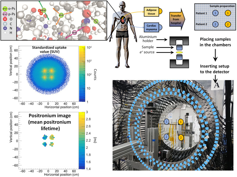
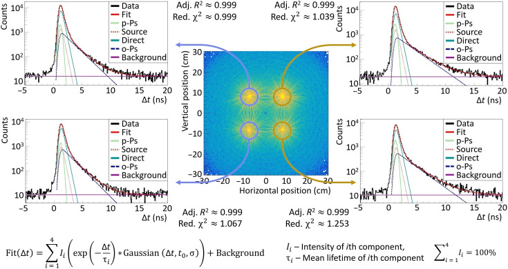
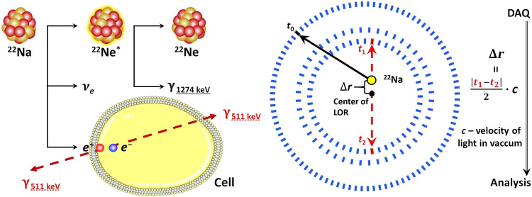
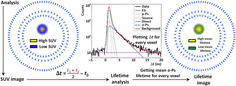
In vivo assessment of cancer and precise location of altered tissues at initial stages of molecular disorders are important diagnostic challenges. Positronium is copiously formed in the free molecular spaces in the patient’s body during positron emission tomography (PET). The positronium properties vary according to the size of inter- and intramolecular voids and the concentration of molecules in them such as, e.g., molecular oxygen, O; therefore, positronium imaging may provide information about disease progression during the initial stages of molecular alterations. Current PET systems do not allow acquisition of positronium images. This study presents a new method that enables positronium imaging by simultaneous registration of annihilation photons and deexcitation photons from pharmaceuticals labeled with radionuclides. The first positronium imaging of a phantom built from cardiac myxoma and adipose tissue is demonstrated. It is anticipated that positronium imaging will substantially enhance the specificity of PET diagnostics.
在分子疾病初始阶段对癌症进行体内评估以及精确确定病变组织的位置是重要的诊断挑战。在正电子发射断层扫描(PET)期间,正电子素在患者体内的自由分子空间中大量形成。正电子素的性质根据分子间和分子内空隙的大小以及其中分子(如分子氧O)的浓度而变化;因此,正电子素成像可以提供有关分子改变初始阶段疾病进展的信息。目前的PET系统不允许采集正电子素图像。本研究提出了一种新方法,通过同时记录来自放射性核素标记药物的湮没光子和去激发光子来实现正电子素成像。展示了由心脏黏液瘤和脂肪组织构建的模型的首次正电子素成像。预计正电子素成像将大大提高PET诊断的特异性。