Unit of Experimental Medicine and Clinical Pathology, Department of Clinical and Biological Sciences, University of Turin, Italy.
Department of Health Sciences and Interdisciplinary Research Centre for Autoimmune Diseases, University Amedeo Avogadro of East Piedmont, Novara, Italy.
Cell Mol Gastroenterol Hepatol. 2022;13(2):459-482. doi: 10.1016/j.jcmgh.2021.10.002. Epub 2021 Oct 14.
BACKGROUND & AIMS: Hypoxia and hypoxia-inducible factors (HIFs) are involved in chronic liver disease progression. We previously showed that hepatocyte HIF-2α activation contributed significantly to nonalcoholic fatty liver disease progression in experimental animals and human patients. In this study, using an appropriate genetic murine model, we mechanistically investigated the involvement of hepatocyte HIF-2α in experimental nonalcoholic steatohepatitis (NASH)-related carcinogenesis.
The role of HIF-2α was investigated by morphologic, cellular, and molecular biology approaches in the following: (1) mice carrying hepatocyte-specific deletion of HIF-2α (HIF-2α mice) undergoing a NASH-related protocol of hepatocarcinogenesis; (2) HepG2 cells stably transfected to overexpress HIF-2α; and (3) liver specimens from NASH patients with hepatocellular carcinoma.
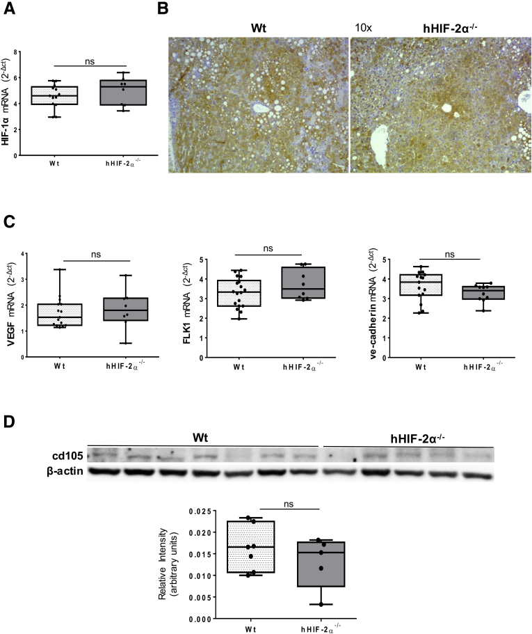
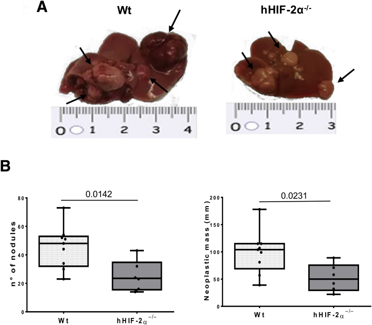
Mice carrying hepatocyte-specific deletion of HIF-2α (hHIF-2α) showed a significant decrease in the volume and number of liver tumors compared with wild-type littermates. These effects did not involve HIF-1α changes and were associated with a decrease of cell proliferation markers proliferating cell nuclear antigen and Ki67. In both human and rodent nonalcoholic fatty liver disease-related tumors, HIF-2α levels were strictly associated with hepatocyte production of SerpinB3, a mediator previously shown to stimulate liver cancer cell proliferation through the Hippo/Yes-associated protein (YAP)/c-Myc pathway. Consistently, we observed positive correlations between the transcripts of HIF-2α, YAP, and c-Myc in individual hepatocellular carcinoma tumor masses, while HIF-2α deletion down-modulated c-Myc and YAP expression without affecting extracellular signal-regulated kinase 1/2, c-Jun N-terminal kinase, and AKT-dependent signaling. In vitro data confirmed that HIF-2α overexpression induced HepG2 cell proliferation through YAP-mediated mechanisms.
These results indicate that the activation of HIF-2α in hepatocytes has a critical role in liver carcinogenesis during NASH progression, suggesting that HIF-2α-blocking agents may serve as novel putative therapeutic tools.
缺氧和缺氧诱导因子(HIFs)参与慢性肝病的进展。我们之前的研究表明,肝细胞 HIF-2α 的激活对实验动物和人类患者的非酒精性脂肪性肝病的进展有重要贡献。在这项研究中,我们使用适当的遗传小鼠模型,从机制上研究了肝细胞 HIF-2α 在实验性非酒精性脂肪性肝炎(NASH)相关致癌中的作用。
通过形态学、细胞和分子生物学方法研究了 HIF-2α 的作用,包括:(1)进行 NASH 相关肝癌发生方案的肝细胞特异性 HIF-2α 缺失(HIF-2α 小鼠)的小鼠;(2)稳定转染过表达 HIF-2α 的 HepG2 细胞;和(3)伴有肝细胞癌的 NASH 患者的肝组织标本。
与野生型同窝仔相比,携带肝细胞特异性 HIF-2α 缺失(hHIF-2α)的小鼠的肝肿瘤体积和数量明显减少。这些作用不涉及 HIF-1α 的变化,并与细胞增殖标志物增殖细胞核抗原和 Ki67 的减少有关。在人和啮齿动物的非酒精性脂肪性肝病相关肿瘤中,HIF-2α 水平与 SerpinB3 的肝细胞产生严格相关,SerpinB3 是一种先前被证明通过 Hippo/Yes 相关蛋白(YAP)/c-Myc 通路刺激肝癌细胞增殖的介质。一致地,我们观察到单个肝癌肿瘤质量中 HIF-2α、YAP 和 c-Myc 的转录物之间存在正相关,而 HIF-2α 的缺失下调了 c-Myc 和 YAP 的表达,而不影响细胞外信号调节激酶 1/2、c-Jun N-末端激酶和 AKT 依赖性信号。体外数据证实,HIF-2α 的过表达通过 YAP 介导的机制诱导 HepG2 细胞增殖。
这些结果表明,HIF-2α 在肝细胞中的激活在 NASH 进展过程中的肝致癌中起着关键作用,这表明 HIF-2α 阻断剂可能作为新的潜在治疗工具。