School of Life and Environmental Sciences, Charles Perkins Centre and Sydney Institute for Infectious Diseases, The University of Sydney, Sydney, NSW, 2006, Australia.
School of Life and Environmental Sciences, School of Medical Sciences, Charles Perkins Centre and Sydney Mass Spectrometry, The University of Sydney, Sydney, NSW, 2006, Australia.
J Neuroinflammation. 2021 Oct 16;18(1):237. doi: 10.1186/s12974-021-02277-x.
Type I interferons (IFN-I) are key responders to central nervous system infection and injury and are also increased in common neurodegenerative diseases. Their effects are primarily mediated via transcriptional regulation of several hundred interferon-regulated genes. In addition, IFN-I activate several kinases including members of the MAPK and PI3K families. Yet, how changes to the global protein phosphoproteome contribute to the cellular response to IFN-I is unknown.
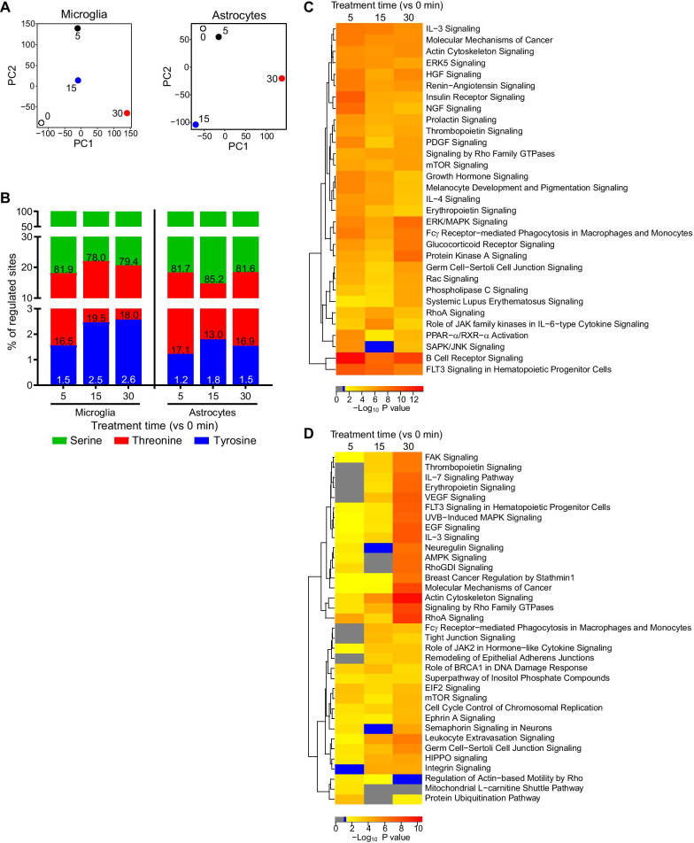
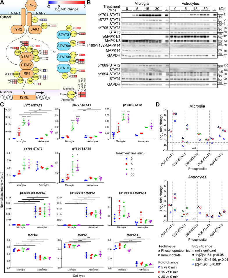
The cerebral phosphoproteome of mice with brain-targeted chronic production of the IFN-I, IFN-α, was obtained. Changes in phosphorylation were analyzed by ontology and pathway analysis and kinase enrichment predictions. These were verified by phenotypic analysis, immunohistochemistry and immunoblots. In addition, primary murine microglia and astrocytes, the brain's primary IFN-I-responding cells, were acutely treated with IFN-α and the global phosphoproteome was similarly analyzed.
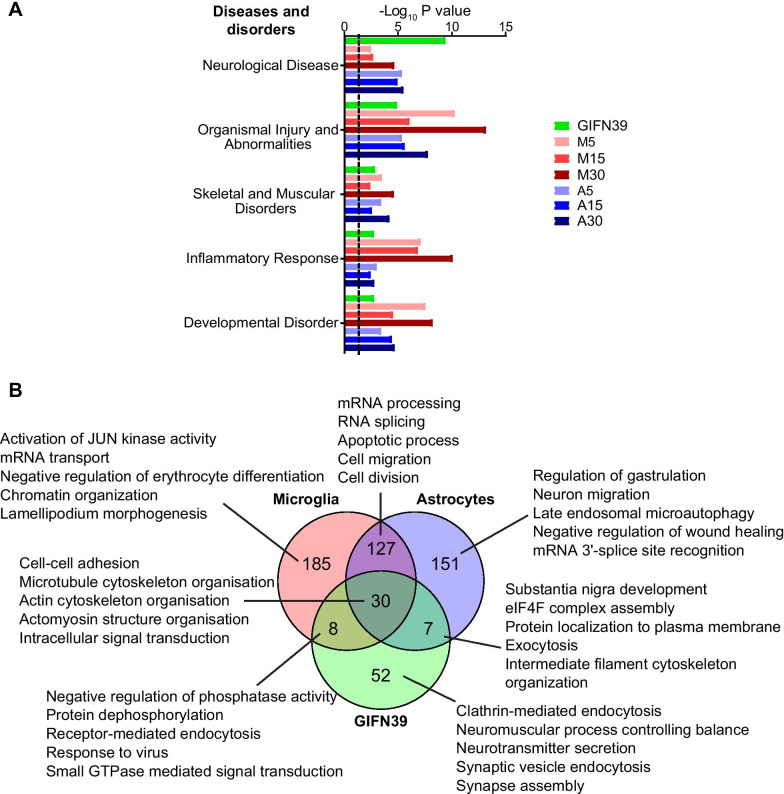
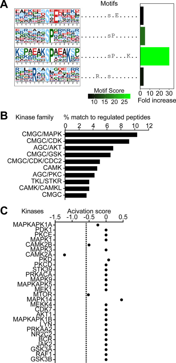
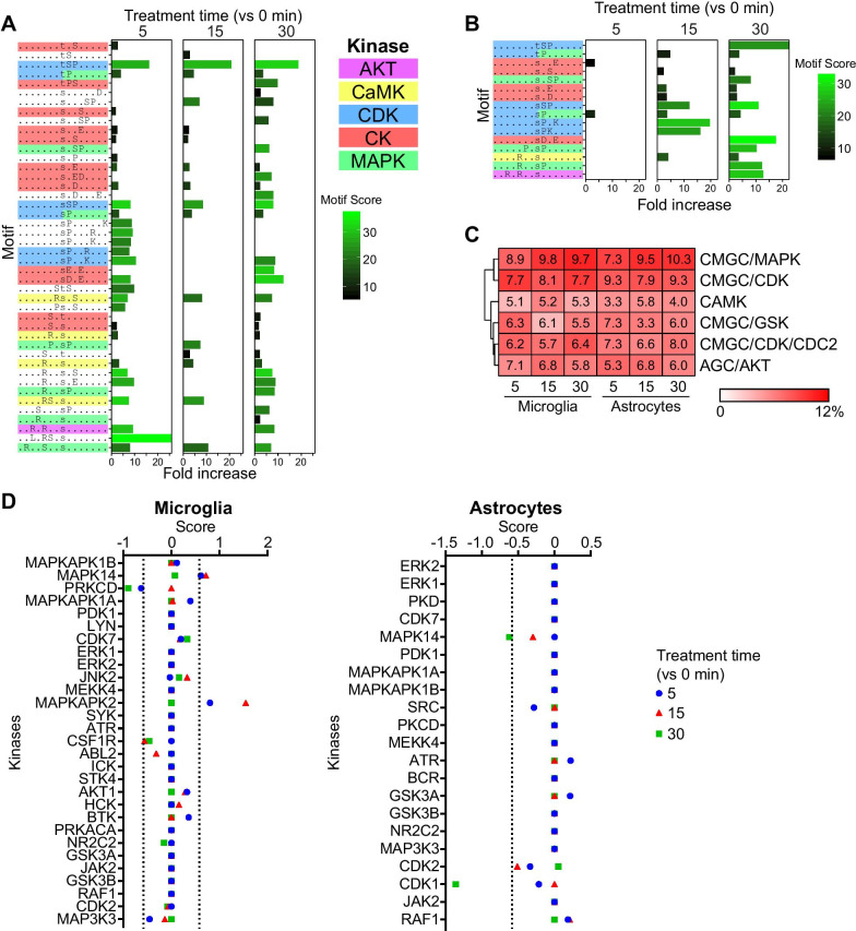
We identified widespread protein phosphorylation as a novel mechanism by which IFN-I mediate their effects. In our mouse model for IFN-I-induced neurodegeneration, protein phosphorylation, rather than the proteome, aligned with the clinical hallmarks and pathological outcome, including impaired development, motor dysfunction and seizures. In vitro experiments revealed extensive and rapid IFN-I-induced protein phosphorylation in microglia and astrocytes. Response to acute IFN-I stimulation was independent of gene expression and mediated by a small number of kinase families. The changes in the phosphoproteome affected a diverse range of cellular processes and functional analysis suggested that this response induced an immediate reactive state and prepared cells for subsequent transcriptional responses.
Our studies reveal a hitherto unappreciated role for changes in the protein phosphorylation landscape in cellular responses to IFN-I and thus provide insights for novel diagnostic and therapeutic strategies for neurological diseases caused by IFN-I.
I 型干扰素 (IFN-I) 是中枢神经系统感染和损伤的主要反应者,也是常见神经退行性疾病中增加的。它们的作用主要通过几种几百个干扰素调节基因的转录调节来介导。此外,IFN-I 激活几种激酶,包括 MAPK 和 PI3K 家族的成员。然而,细胞对 IFN-I 反应的全局蛋白磷酸化组变化如何尚不清楚。
获得了大脑靶向慢性产生 IFN-I、IFN-α 的小鼠大脑磷酸蛋白质组。通过本体论和途径分析以及激酶富集预测分析磷酸化的变化。通过表型分析、免疫组织化学和免疫印迹进行了验证。此外,急性用 IFN-α处理原代小鼠小胶质细胞和星形胶质细胞,即大脑中主要的 IFN-I 反应细胞,并对其进行了全蛋白磷酸化组分析。
我们发现广泛的蛋白质磷酸化是 IFN-I 介导其作用的新机制。在我们的 IFN-I 诱导神经退行性变的小鼠模型中,蛋白质磷酸化而不是蛋白质组与临床特征和病理结果一致,包括发育受损、运动功能障碍和癫痫发作。体外实验显示 IFN-I 诱导的小胶质细胞和星形胶质细胞中广泛而迅速的蛋白质磷酸化。对急性 IFN-I 刺激的反应独立于基因表达,并由少数几个激酶家族介导。磷酸蛋白质组的变化影响了广泛的细胞过程,功能分析表明该反应诱导了即时反应状态,并为随后的转录反应做好了准备。
我们的研究揭示了蛋白质磷酸化图谱变化在细胞对 IFN-I 反应中的一个以前未被认识的作用,从而为 IFN-I 引起的神经疾病的新型诊断和治疗策略提供了新的见解。