Department of Orthopaedic Surgery, Kobe University Graduate School of Medicine, 7-5-2, Kusunoki-cho, Chuo-ku, Kobe, 650-0017, Japan.
Laboratory of Biomaterials, Institute for Frontier Life and Medical Sciences, Kyoto University, 53 Kawara-cho Shogoin, Sakyo-ku, Kyoto, 606-8507, Japan.
J Orthop Surg Res. 2021 Oct 16;16(1):605. doi: 10.1186/s13018-021-02771-1.
Excellent outcomes of arthroscopic rotator cuff repair for small and medium tears have been recently reported. However, re-tears after surgery have been a common complication after surgical repair of large and massive rotator cuff tears and often occur in early postoperative phase. It was previously reported that basic fibroblast growth factor and platelet-rich plasma enhanced rotator cuff tear healing. We hypothesized that this combined therapy could enhance rotator cuff healing after rotator cuff repair in a rat model. This study aimed to evaluate the efficacy of combined therapy of platelet-rich plasma and basic fibroblast growth factor with gelatin-hydrogel sheet.
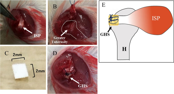
To create a rotator cuff defect, the infraspinatus tendon of Sprague Dawley rat was resected from the greater tuberosity. The infraspinatus tendons were repaired and covered with gelatin-hydrogel sheet impregnated with PBS (control group), basic fibroblast growth factor (bFGF group), platelet-rich plasma (PRP group), or both basic fibroblast growth factor and platelet-rich plasma (combined group). Histological examinations were conducted using hematoxylin and eosin, safranin O, and immunofluorescence staining, such as Isolectin B4, type II collagen at 2 weeks postoperatively. For mechanical analysis, ultimate failure load of the tendon-humeral head complex was evaluated at 6 weeks postoperatively.
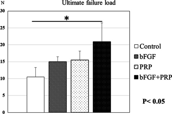
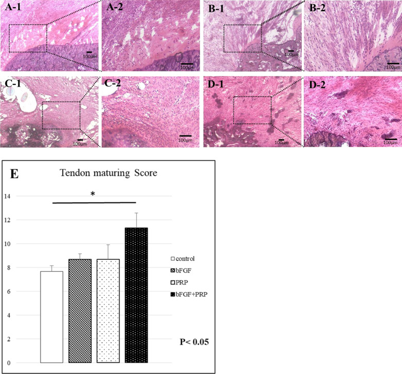
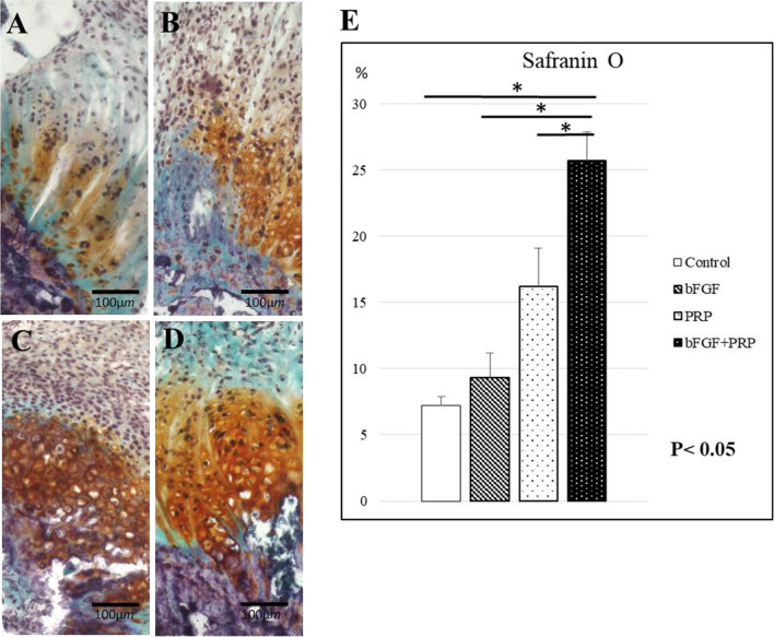
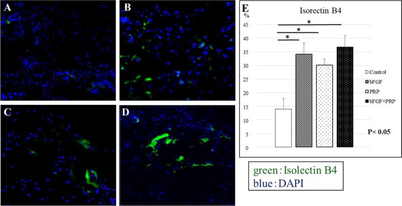
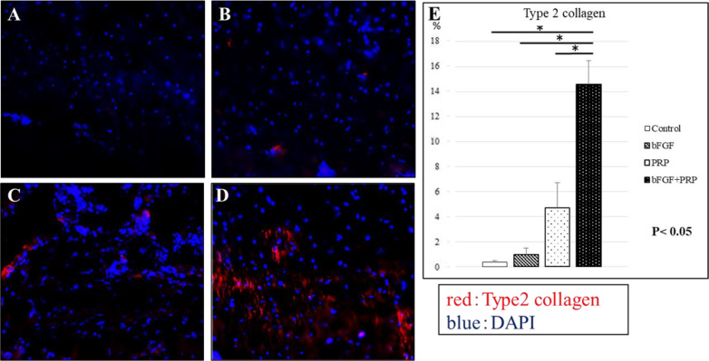
In the hematoxylin and eosin staining, the tendon maturing score of the combined group was higher than that of the control group at postoperative 2 weeks. In the safranin O staining, stronger proteoglycan staining was observed in the combined group compared with the other groups at postoperative 2 weeks. Vascular staining with isolectin B4 in 3 treatment groups was significantly higher than that in the control group. Type II collagen expression in the combined group was significantly higher than those in the other groups. The ultimate failure load of the combined group was significantly higher than that of the control group.
Combined therapy of basic fibroblast growth factor and platelet-rich plasma promoted angiogenesis, tendon maturing and fibrocartilage regeneration at the enthesis, which could enhance the mechanical strength. It was suggested that combined basic fibroblast growth factor and platelet-rich plasma might enhance both tendon and bone-tendon junction healing, and basic fibroblast growth factor and platelet-rich plasma might be synergistic.
最近有报道称,关节镜下修复小、中撕裂的肩袖的效果非常好。然而,对于大、巨大肩袖撕裂的手术修复后再次撕裂仍然是一种常见的并发症,而且常常发生在术后早期。此前有研究报道称,碱性成纤维细胞生长因子和富血小板血浆可以促进肩袖撕裂的愈合。我们假设这种联合治疗可以在大鼠模型中增强肩袖修复后的肩袖愈合。本研究旨在评估富血小板血浆和碱性成纤维细胞生长因子与明胶-水凝胶片联合治疗的效果。
为了制造肩袖缺损,从肩胛冈突上切除冈下肌腱。冈下肌腱修复后,用明胶-水凝胶片覆盖,该明胶-水凝胶片用 PBS(对照组)、碱性成纤维细胞生长因子(bFGF 组)、富血小板血浆(PRP 组)或碱性成纤维细胞生长因子和富血小板血浆(联合组)浸渍。术后 2 周,通过苏木精和伊红、番红 O 和免疫荧光染色(如异硫氰酸荧光素 B4、II 型胶原)进行组织学检查。术后 6 周,评估腱-肱骨头复合体的极限失效负荷。
在苏木精和伊红染色中,联合组在术后 2 周时的肌腱成熟评分高于对照组。在番红 O 染色中,联合组在术后 2 周时的蛋白聚糖染色比其他组更强。3 种治疗组的异硫氰酸荧光素 B4 血管染色明显高于对照组。联合组的 II 型胶原表达明显高于其他组。联合组的极限失效负荷明显高于对照组。
碱性成纤维细胞生长因子和富血小板血浆联合治疗促进了腱-骨结合处的血管生成、肌腱成熟和纤维软骨再生,从而增强了机械强度。这表明联合应用碱性成纤维细胞生长因子和富血小板血浆可能增强腱和腱-骨结合部的愈合,且碱性成纤维细胞生长因子和富血小板血浆可能具有协同作用。