Planek Maria Isabel Camara, Ruge Max, Du Fay de Lavallaz Jeanne M, Kyung Stella B, Gomez Joanne Michelle D, Suboc Tisha M, Williams Kim A, Volgman Annabelle Santos, Simmons J Alan, Rao Anupama K
Department of Internal Medicine, Rush University Medical Center, Chicago, IL, United States of America.
Department of Internal Medicine, Thomas Jefferson University, Philadelphia, PA, United States of America.
Am Heart J Plus. 2021 Nov;11:100052. doi: 10.1016/j.ahjo.2021.100052. Epub 2021 Oct 13.
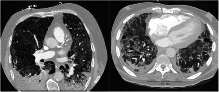
Chest computed tomography (chest CT) is routinely obtained to assess disease severity in COVID-19. While pulmonary findings are well-described in COVID-19, the implications of cardiovascular findings are less well understood. We evaluated the impact of cardiovascular findings on chest CT on the adverse composite outcome (ACO) of hospitalized COVID-19 patients.
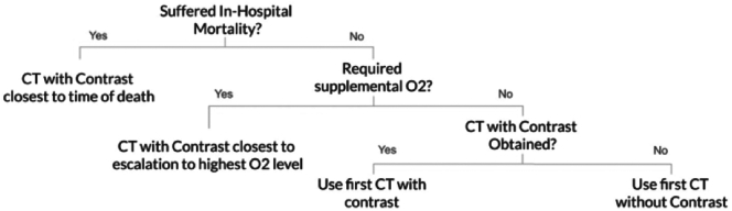
SETTING/PARTICIPANTS: 245 COVID-19 patients who underwent chest CT at Rush University Health System were included.
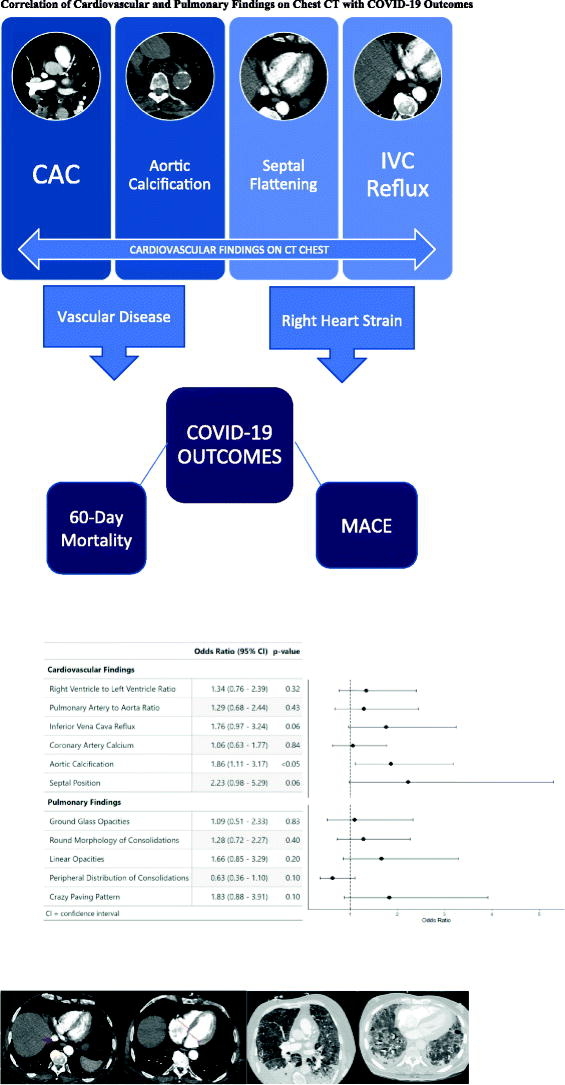
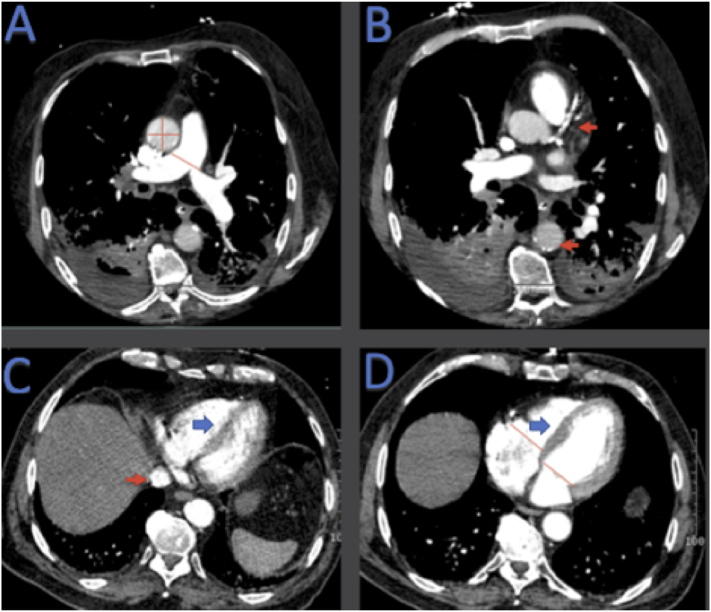
Cardiovascular findings, including coronary artery calcification (CAC), aortic calcification, signs of right ventricular strain [right ventricular to left ventricular diameter ratio, pulmonary artery to aorta diameter ratio, interventricular septal position, and inferior vena cava (IVC) reflux], were measured by trained physicians.
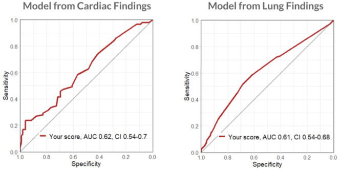
INTERVENTIONS/MAIN OUTCOME MEASURES: These findings, along with pulmonary findings, were analyzed using univariable logistic analysis to determine the risk of ACO defined as intensive care admission, need for non-invasive positive pressure ventilation, intubation, in-hospital and 60-day mortality. Secondary endpoints included individual components of the ACO.
Aortic calcification was independently associated with an increased risk of the ACO (odds ratio 1.86, 95% confidence interval (1.11-3.17) < 0.05). Aortic calcification, CAC, abnormal septal position, or IVC reflux of contrast were all significantly associated with 60-day mortality and major adverse cardiovascular events. IVC reflux was associated with in-hospital mortality ( = 0.005).
Incidental cardiovascular findings on chest CT are clinically important imaging markers in COVID-19. It is important to ascertain and routinely report cardiovascular findings on CT imaging of COVID-19 patients as they have potential to identify high risk patients.
胸部计算机断层扫描(胸部CT)常用于评估新型冠状病毒肺炎(COVID-19)患者的疾病严重程度。虽然COVID-19的肺部表现已有详细描述,但心血管系统表现的意义尚不清楚。我们评估了胸部CT上的心血管系统表现对住院COVID-19患者不良复合结局(ACO)的影响。
研究地点/参与者:纳入了245例在拉什大学医疗系统接受胸部CT检查的COVID-19患者。
由经过培训的医生测量心血管系统表现,包括冠状动脉钙化(CAC)、主动脉钙化、右心室应变迹象[右心室与左心室直径比、肺动脉与主动脉直径比、室间隔位置和下腔静脉(IVC)反流]。
干预措施/主要结局指标:这些表现以及肺部表现通过单变量逻辑分析进行分析,以确定ACO的风险,ACO定义为重症监护病房入住、无创正压通气需求、插管、住院和60天死亡率。次要终点包括ACO的各个组成部分。
主动脉钙化与ACO风险增加独立相关(比值比1.86,95%置信区间(1.11-3.17)<0.05)。主动脉钙化、CAC、室间隔位置异常或IVC造影剂反流均与60天死亡率和主要不良心血管事件显著相关。IVC反流与住院死亡率相关(=0.005)。
胸部CT上偶然发现的心血管系统表现在COVID-19中是重要的临床影像学标志物。在COVID-19患者的CT成像中确定并常规报告心血管系统表现很重要,因为它们有可能识别高危患者。