Department of Diagnostic and Interventional Radiology, Faculty of Medicine and University Hospital Cologne, University Cologne, Cologne, Germany.
Department of Radiology, Helios Dr. Horst Schmidt Kliniken Wiesbaden, Wiesbaden, Germany.
PLoS One. 2020 Dec 23;15(12):e0244267. doi: 10.1371/journal.pone.0244267. eCollection 2020.
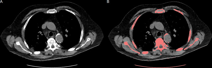
Cardiovascular comorbidity anticipates poor prognosis of SARS-CoV-2 disease (COVID-19) and correlates with the systemic atherosclerotic transformation of the arterial vessels. The amount of aortic wall calcification (AWC) can be estimated on low-dose chest CT. We suggest quantification of AWC on the low-dose chest CT, which is initially performed for the diagnosis of COVID-19, to screen for patients at risk of severe COVID-19.
Seventy consecutive patients (46 in center 1, 24 in center 2) with parallel low-dose chest CT and positive RT-PCR for SARS-CoV-2 were included in our multi-center, multi-vendor study. The outcome was rated moderate (no hospitalization, hospitalization) and severe (ICU, tracheal intubation, death), the latter implying a requirement for intensive care treatment. The amount of AWC was quantified with the CT vendor's software.
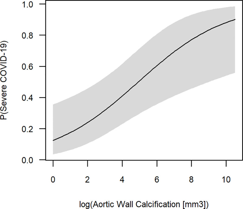
Of 70 included patients, 38 developed a moderate, and 32 a severe COVID-19. The average volume of AWC was significantly higher throughout the subgroup with severe COVID-19, when compared to moderate cases (771.7 mm3 (Q1 = 49.8 mm3, Q3 = 3065.5 mm3) vs. 0 mm3 (Q1 = 0 mm3, Q3 = 57.3 mm3)). Within multivariate regression analysis, including AWC, patient age and sex, as well as a cardiovascular comorbidity score, the volume of AWC was the only significant regressor for severe COVID-19 (p = 0.004). For AWC > 3000 mm3, the logistic regression predicts risk for a severe progression of 0.78. If there are no visually detectable AWC risk for severe progression is 0.13, only.
AWC seems to be an independent biomarker for the prediction of severe progression and intensive care treatment of COVID-19 already at the time of patient admission to the hospital; verification in a larger multi-center, multi-vendor study is desired.
心血管合并症预示着严重急性呼吸综合征冠状病毒 2 型疾病(COVID-19)的预后不良,并与动脉血管的系统性动脉粥样硬化转化相关。主动脉壁钙化(AWC)的量可以通过低剂量胸部 CT 来估计。我们建议在最初用于 COVID-19 诊断的低剂量胸部 CT 上定量 AWC,以筛选出 COVID-19 高危患者。
我们进行了一项多中心、多供应商的研究,共纳入了 70 名连续的 COVID-19 患者(中心 1 组 46 名,中心 2 组 24 名),这些患者均并行低剂量胸部 CT 检查,并且 SARS-CoV-2 的 RT-PCR 检测为阳性。结果被评为中度(无住院、住院)和重度(重症监护病房、气管插管、死亡),后者意味着需要重症监护治疗。AWC 的量使用 CT 供应商的软件进行定量。
在 70 名纳入的患者中,38 名患者发展为中度 COVID-19,32 名患者发展为重度 COVID-19。与中度病例相比,重度 COVID-19 患者的 AWC 总体积明显更高(771.7 mm3(Q1=49.8 mm3,Q3=3065.5 mm3)vs. 0 mm3(Q1=0 mm3,Q3=57.3 mm3))。在包括 AWC、患者年龄和性别以及心血管合并症评分的多变量回归分析中,AWC 是重度 COVID-19 的唯一显著预测因子(p=0.004)。对于 AWC>3000 mm3,逻辑回归预测重度进展的风险为 0.78。如果没有明显的 AWC 风险,则预测重度进展的风险为 0.13,仅为前者的 1/7。
AWC 似乎是预测 COVID-19 严重进展和重症监护治疗的独立生物标志物,可在患者入院时使用;需要在更大的多中心、多供应商研究中进行验证。